WCNR-11 - 11th World Conference on Neutron Radiography

Australia/Sydney
Lighthouse Gallery (Australian National Maritime Museum)

Lighthouse Gallery

Australian National Maritime Museum

2 Murray Street, Sydney NSW 2000
Ulf Garbe (ANSTO)
    • 15:30 18:00
      Board Meeting
    • 18:00 20:30
      Reception and Welcome Meeting
    • 08:00 08:45
      Registration
    • 08:45 09:00
      Opening
    • 09:00 10:50
      Speaker Sessions and Seminars
      • 09:00
        Pulsed neutron imaging 50m

        Pulsed neutron sources are useful for imaging since they give different information compared with steady reactor sources [1,2]. At the pulsed neutron source, we can easily use the time-of-flight method to analyze the neutron energy and it enables us to obtain position dependent transmission data as a function of energy with high energy resolution. The transmission spectrum is affected by neutron interaction cross section, and by analyzing the energy dependency of the transmission we can deduced quantitative information of an object. ‘Quantitative evaluation’ is one of the most important characteristics of the pulsed neutron imaging.
        There are various kinds of neutron interactions with matters. Coherent scattering is most important interaction to deduce the crystallographic information. We can get information on crystal phase, crystallite size, preferred orientation (texture) [3], and strain [4,5]. Micro-strain information will be included in the Bragg edge transmission [6]. Incoherent scattering does not have strong energy dependency. One of applications is to use the increase of low energy cross section of hydrogen, and the hump of cross section due to oscillation of hydrogen atom in a metal hydride [7]. Another feature of the neutron cross section is resonance peaks at higher energy. Information on dynamics of a peculiar element was studied [8], temperature was measured [9] and elemental distribution in a sample was obtained [10]. Other special feature of neutrons is magnetic interaction. By using the magnetic interaction combined with energy dependent transmission, we can evaluate the absolute value of the magnetic field [7, 11].
        Material analysis and cultural heritage researches are performed by using pulsed neutron sources, and further development of data analysis is still ongoing. In the presentation, the pulsed neutron imaging method and its applications are presented.  

        References
        [1] Y. Kiyanagi and H. Iwasa, Proceedings of the Fifth International Symposium on Advanced Nuclear Energy Research—Neutron as Microscope Probes, JAERI-M 93-228, Ibaraki, Japan, 10-12 Mar 1993, Vol.2, pp.796-801
        [2] Y. Kiyanagi, T. Kamiyama, H. Iwasa and F. Hiraga, Key Engineering Materials, Vol.270-273, pp.1371-1375, (2004)
        [3] H. Sato, T. Kamiyama, Y. Kiyanagi, Materials Transactions, Vol.52, No.6, pp.1294-1302, (2011)
        [4] J. R. Santisteban, L. Edwards, A. Steuwer and P. J. Withers, J. Appl. Cryst. 34, (2001) 289.
        [5] J. R. Santisteban, L. Edwards, M. E. Fizpatrick, A. Steuwer, P. J. Withers, Appl. Phys. A 74, (2002) S1433.
        [6] T. Kamiyama, K. Iwase, H. Sato, S. Harjo, T. Ito, S. Takata, K. Aizawa, Y. Kiyanagi, Physics Procedia 88 ( 2017) 50 – 57.
        [7] Y. Kiyanagi, H. Sato, T. Kamiyama and T. Shinohara, J. Physics, Conference Series 340, 012010 (2012).
        [8] K. Kaneko, T. Kamiyama, Y. Kiyanagi, T. Sakuma, S. Ikeda, J. Physics and Chemistry of Solids, Vol.60, pp.1499-1502, (1999).
        [9] K. Tokuda, T. Kamiyama, Y. Kiyanagi, R. Moreh and S. Ikeda, J. J. Applied Physics, vol.40, No.3A, pp.1504-1507, (2001).
        [10] H. Sato, T. Kamiyama, Y. Kiyanagi, S. Ikeda, Nucl. Instr. Meth., Physics Research, A600, pp.135-138, (2009).
        [11] T. Shinohara, et al., J. Physics: Conf. Series 862 (2017) 012025.
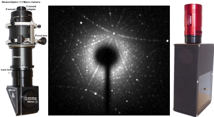
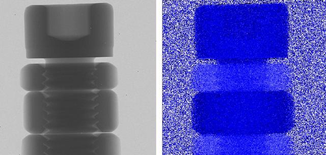
        Speaker: Yoshiaki Kiyanagi (Nagoya University)
      • 09:50
        Imaging at the Spallation Neutron Source: Opportunities and Challenges 20m

        Over the past few years, several wavelength-dependent neutron imaging capabilities have been developed at spallation neutron sources such as RADEN at J-PARC and IMAT at ISIS. At the Spallation Neutron Source of Oak Ridge National Laboratory, wavelength-dependent experiments are ongoing, and a temporary imaging capability is being planned at the Spallation Neutrons and Pressure Diffractometer (SNAP), beamline 3) instrument. A design of this new imaging capability is presented. The facility will be equipped with exchangeable apertures optimized for cold, thermal and epithermal neutrons, respectively. A dedicated sample area (for 2D and 3D data acquisition) and in-house event mode microchannel plate (MCP) detector are currently being developed as part of this project.
        Recently, the team has measured crystalline structures (using cold neutrons) and isotopic content (using epithermal neutrons) in superalloys and nuclear fuel material, respectively. We present the characterization of additively manufactured (AM) Inconel 718 using wavelength-dependent radiography, the so-called Bragg edge imaging technique, diffraction and modeling. This dual-modality capability combined with modeling provides unique information about the crystalline

        Speaker: Hassina Bilheux (Oak Ridge National Laboratory)
      • 10:10
        Wavelength-resolved neutron imaging on IMAT 20m

        The ‘IMAT’ instrument, which specializes in Imaging and MATerials science, is now well into its commissioning phase. The basic performance parameters for white-beam tomography and energy-dispersive neutron imaging have been determined [ 1 ] and the instrument is currently being prepared for user operation [ 2 ]. Here we report on the evaluation of the wavelength-resolving imaging options on IMAT, including pink-beam imaging using disk choppers and energy-dispersive Bragg edge imaging using time-resolving detectors. These time-of-flight techniques enable image contrast enhancement and mapping of structure properties. We will review the recent infrastructure installations and software developments that have been undertaken to take advantage of these techniques, making the facility ready for applications in a diverse range of disciplines such as engineering material science, battery research, earth science and cultural heritage.

        [ 1 ] T. Minniti, et al., Nucl. Instr. Methods A 888 (2018) 184.
        [ 2 ] W. Kockelmann et al., J. Imaging 4 (2018) 47.

        Speaker: Dr Winfried Kockelmann (STFC Rutherford Appleton Laboratory )
      • 10:30
        Current Status of The Energy-Resolved Neutron Imaging System, RADEN, at J-PARC MLF 20m

        The world’s first pulsed neutron imaging instrument dedicated to energy-resolved neutron imaging experiments, named RADEN, was constructed at beam port 22 in the Materials and Life Science Experimental Facility (MLF) of J-PARC [1]. This instrument is designed to conduct state-of-the-art energy-resolved neutron imaging, such as Bragg-edge imaging, resonance absorption imaging, and pulsed polarized neutron imaging, together with conventional/energy-selective neutron radiography and tomography by fully utilizing the high intensity, short-pulsed neutron beam. The construction of RADEN was completed in 2014, and user operation was started from April 2015. By the end of April 2017, the number of conducted proposals reached 72 and about half of the total beam time was utilized by general users.
        Besides the user program, the RADEN instrument group is continuing the technical development and improvement of the instrument so as to conduct more advanced energy-resolved and conventional neutron imaging experiments. To improve the detector performance, we exchanged the optical system of our camera-type detector for increased brightness to achieve fine spatial resolution, and upgraded an event-type detector, the µNID [2], for improved count rate, neutron detection efficiency, and spatial resolution. Also, the collimation system was upgraded and additional slits were introduced into the instrument for better beam shaping and efficient background reduction. The device controlling software has been replaced with a newer version in order to make the interface more user friendly and to provide flexiblity to easily include additional equipment under the control. Regarding the development of new imaging techniques, we have constructed a Talbot-Lau interferometer at the pulsed neutron source for the first time and applied the wavelength-dependent analysis to phase imaging [3]. Moreover, to analyze the spatial distribution of nano-scale structural information, a technique to extract the small-angle scattering contribution in the neutron transmission spectrum using orthogonally-arranged neutron Soller collimators has been developed [4].
        In this presentation, we will report the current status of RADEN along with recent results of the technical development and application studies regarding energy-resolved neutron imaging techniques conducted at RADEN.
        This work was partially supported by the Photon and Quantum Basic Research Coordinated Development Program from MEXT, Japan and the Momose Quantum Beam Phase Imaging Project, ERATO, JST (Grant No. JPMJER1403).
        [1] T. Shinohara et al., J. Phys. Conf. Ser., 746, 012007 (2016).
        [2] J.D. Parker et al., Nucl. Instr. and Meth. A, 726, 155 (2013).
        [3] Y. Seki et al., J. Phys. Soc. Jpn., 86, 044001 (2017).
        [4] Y. Oba et al., J. Appl. Cryst., 50, 334 (2017).

        Speaker: Dr Takenao Shinohara (Japan Atomic Energy Agency)
    • 10:50 11:10
      Morning Tea 20m
    • 11:10 12:30
      Speaker Sessions and Seminars
      • 11:10
        Novel scintillation screen with significantly improved radiation hardness and very high light output 20m

        Novel scintillation screen with significantly improved radiation hardness and very high light output

        Bernhard Walfort1, Christian Grünzweig2, Pavel Trtik2, Manuel Morgano2, Markus Strobl2
        1 RC Tritec AG, Speicherstrasse 60 a, CH-9053 Teufen, Switzerland, walfort@rctritec.com
        2 Paul Scherrer Institut, Laboratory for Neutron Scattering and Imaging (LNS), WBBA 108, CH-5232 Villigen, Switzerland; christian.gruenzweig@psi.ch

        That the bombardment with a high energy radiation has an effect on the material properties is well known since many years [1]. Vacancies or disorder incorporated in the material can have a significant influence on the luminous intensity of scintillation material. In the development of scintillation material for high energy particle detection big efforts have been done within the last years [2]. Actually the highly radiation resistant garnets seem to be the state of the art in that field [2].
        It is known that neutrons affecting, especially the alpha and triton particle originating from the capture reaction, lead to a degradation of scintillator screens regarding the light output versus neutron fluence. Two different kind of scintillators are commonly used for neutron imaging applications: (i) 6LiF/ZnS scintillation screens with very high light output and reasonable resolution and (ii) Gd2O2S:Tb scintillation screens for very high resolution measurements but 10 times less light output.
        Within a 2 years development project with PSI we wanted to understand for the 6LiF/ZnS scintillator type the effects which are responsible for the degradation mechanism and therewith be able to develop a more radiation hard neutron scintillator system. The goal was to not only improve radiation hardness but also to improve the light output in comparison to the traditional 6LiF/ZnS-scintillation screens.
        In this talk the results of this development project, leading to a new type of scintillation screen with significantly improved radiation hardness, but still very high light output, are presented.

        [1] Kurt E. Sickafus, Eugene A. Kotomin, Blas P. Uberuaga “Radiation Effects in Solids” Springer, 2004. ISBN-10 1-4020-5294-4 (PB) .
        [2] P.Lecoq, A.Gektin, M.Korzhik, Scintillation material for detector systems, Springer, 2017, P.408.

        Speaker: Bernhard Walfort
      • 11:30
        Daniel 20m
      • 11:50
        Volume Graphics 40m
    • 12:30 13:30
      Lunch 1h
    • 13:30 15:10
      Speaker Sessions and Seminars
      • 13:30
        Development of energy-selective and element-sensitive imaging using a compact D-D fast neutron generator 20m

        This work is focused on the development of energy-selective techniques using a compact Deuterium-Deuterium (D-D) fast neutron generator. This was done in the context of a custom D-D generator located at the Paul Scherrer Institute which was specifically developed to have a small emitting spot size for transmission imaging purposes [1]. The basis of this study lies in the physics of the D-D fusion reaction: the neutrons produced are quasi-monoenergetic with an energy dependent on emission angle from roughly 2.2 to 2.8 MeV, based on the acceleration voltage limitation of the device. Samples can therefore be imaged at different emission angles corresponding to different neutron energies. Since neutron cross-sections have energy dependence unique to each element (unlike X-rays), this combination of information from different angles can be used in principle to distinguish one element or chemical from another. The inverse can also be performed; instead of determining the content of an unknown sample, measurements of a known and uniform sample can be used to produce cross-section data.

        The first steps of this investigation included a feasibility study of these techniques. Detailed angle-dependent source emission spectra models were created according to different target composition assumptions. These models were used to estimate attenuation vs. angle for several samples of known composition and thickness which have particularly prominent cross-section structures in the energy range of interest (e.g. alumina). Over the full range of emission angles, plastic scintillators were used to measure count rates with sample present, without sample, and with a shadow cone, in order to determine the sample attenuation. This was done with a custom, automated mechanical apparatus around the source. Scatter correction was also implemented based on detailed Monte Carlo simulations of the source and room geometry. The experimental attenuation data were compared with simulations and found to be in good agreement, demonstrating the fundamental feasibility of the approach.

        Ongoing work aims to expand these measurements to include a range of materials which are of interest to industrial or homeland security applications. Furthermore, the next step of performing full tomographic reconstruction at multiple angles is being explored, both with simulations and measurements. The aim is to find the practical capabilities and limitations of determining the presence of materials of interest in samples of unknown composition. The latest results and progress towards this goal will be presented and discussed.


        References
        [1] R. Adams, R. Zboray, H.-M. Prasser - A novel fast-neutron tomography system based on a plastic scintillator array and a compact D–D neutron generator - Applied Radiation and Isotopes, 107:1-7, January 2016.

        Speaker: Mr Benoit Soubelet (ETH Zürich)
      • 13:50
        Recent Advances in Neutron Imaging using a High-Flux Accelerator-Based Neutron Generator 20m

        Neutron imaging systems have been designed and constructed by Phoenix LLC to investigate low density material attributes of composites and other materials where other Non-destructive evaluation, (NDE), methods do not suffice. The first-generation electronic neutron generator was commissioned in 2013 at a United States Army research facility to inspect munitions and other critical defense and aerospace components. A second-generation neutron imaging system has undergone extensive testing at the Phoenix laboratory with an increased total neutron output from an upgraded gaseous deuterium target of 3x10^11 deuterium-deuterium (DD) neutrons/second. This system generates a higher neutron flux at the imaging plane, approximately 1x10^4 n/cm^2-sec, which reduces interrogation time, while maintaining high contrast and low geometric unsharpness. A further optimized system is currently under construction and promises yet even higher neutron output with increased image quality regarding signal to noise, contrast, and resolution. This system is expected to be installed at a production plant in 2018 and will be the first of its kind installed and used in a commercial setting. Phoenix’s technology offers high throughput and image quality for neutron radiographs, like images currently acquired at nuclear reactors, but with greater accessibility, an eased regulatory environment, at a much-reduced cost, and without great environmental or biological hazards. As neutron radiography becomes more accessible due to the increased neutron yield of accelerator-based systems, a wider range of inspection techniques will be possible including digital radiography and computed tomography, with shorter image acquisition times. A description of the Phoenix neutron generator and imaging system, including the beamline, target, moderator and collimator, various detector platforms, and post processing techniques including neutron interaction localization and computed tomography, are demonstrated in this presentation. Neutron radiographs captured with a Phoenix neutron radiography system will be presented for both analog and digital based formats.

        Keywords:

        Neutron, radiography, munitions, artillery, aerospace, imaging, direct radiography, computed radiography, computed tomography

        Michael Taylor
        Phoenix LLC (www.phoenixwi.com)
        2555 Industrial Drive
        Monona, WI 53713 USA
        608/210-3060
        michael.taylor@phoenixwi.com

        Speaker: Mr Michael Taylor (Phoenix LLC)
      • 14:10
        HIGH RESOLUTION HIGH ENERGY NEUTRON COMPUTED TOMOGRAPHY AT LANSCE-WNR 20m

        It has long been recognized that neutrons can compliment x-rays for imaging. This is due to their very different attenuation characteristics based on nuclear cross-section, which allows imaging of low Z materials through higher Z materials. Additionally one can use energy dependent Time of Flight (ToF) imaging to exploit phenomenon like nuclear resonances for isotope and element specific imaging. The Los Alamos Neutron Science Center (LANSCE) accelerator is an 800 MeV proton linear accelerator which supplies protons to a range of missions including two spallation neutron targets, one moderated (water and liquid hydrogen) and one unmoderated. This combination of targets provides flight paths which have cold, thermal to epi-thermal and fast neutron energy ranges. In addition the proton pulse structure of the LANSCE accelerator provides neutron pulse lengths of < 270ns for the thermal/cold flight paths and < 1ns for the fast flight paths. These pulse lengths allow for energy discrimination from eV to ~100 MeV.
        Over the last 6 years there has been significant renewed interest in utilizing this source for neutron imaging as a complement to existing x-ray and proton imaging capabilities at LANL. To this end thermal to epi-thermal integrated and ToF imaging (2D radiography and Computed Tomography or CT) have been established and cold neutron propogation based phase contrast imaging has been demonstrated. Finally, significant work has been put into developing a fast neutron imaging capability with the goal of reaching sub mm resolution on objects with an integrated density > 200 g/cm2 and a CT scan time of less than 12 hrs. Fast neutron imaging at high resolution is an area with relatively sparse development due to a lack of available high intensity sources. This talk focuses on advances made in fast neutron imaging at LANSCE-WNR over the last 4 years including flight path modifications, scintillator development and detector testing. Results are shown for a range of scintillators, flat panel detectors and lens coupled camera systems. In addition energy discriminating Time of Flight images from 2 to 60 MeV are shown. Imaging results are shown on imaging quality indicators, a range of industrial parts (cracking, casting voids, etc) and on fossils of various sizes. Where available x-ray CT results are shown on the same parts to demonstrate the pros and cons of fast neutron imaging. Finally, ongoing work and outlook for continued improvement in fast neutron imaging will be discussed.

        Speaker: James Hunter (Los Alamos National Lab)
      • 14:30
        Evaluation of micro-strain, dislocation density and crystallite size from broadening of multiple Bragg-edges observed by pulsed neutron transmission imaging 20m

        It is recognized that Bragg-edge neutron transmission method can deduce crystal structure, crystalline phase, crystallographic texture, crystallite size (from the primary extinction effect) and macro-strain in the imaging mode. In this study, further material information, micro-strain, dislocation density and crystallite size, were deduced by broadening analysis of multiple Bragg-edges.
         So far, we have investigated that Bragg-edge broadening (FWHM of d-spacing distribution) is same as diffraction peak FWHM [1], and proportional to ferrite/martensite ratio and the Vickers hardness [2]. However, the FWHM can be separated to the crystallite size component and the micro-strain component relating to dislocation density [3]. In addition, the dislocation density is very important information for material strength characterizations. For this reason, we tried to separate these broadening components, and deduce micro-strain, crystallite size and dislocation density by using the Williamson-Hall (WH) method. The WH method needs line-broadening information of various diffraction indices.
         Pulsed neutron transmission and diffraction experiment [1,4] was performed at J-PARC MLF BL19 “TAKUMI”. During a tensile test of a low-carbon ferritic steel plate, both data of transmission (by 256-pixels Li-6 glass-scintillator detector) and diffraction (by TAKUMI) were measured. As a result, Bragg-edges and diffraction peaks of various diffraction indices were obtained.
         We firstly checked the classical WH (cWH) plots [3] of both transmission data and diffraction data. This shows relation between Bragg-edge broadenings and diffraction peak broadenings for various diffraction indices. As a result, it was confirmed that Bragg-edge broadenings corresponded to diffraction peak broadenings. In addition, it was correctly observed that the cWH plots did not have linearity due to the anisotropic elasticity. Thus, Bragg-edge broadening is consistent with diffraction peak broadening for multiple diffraction indices. This means that the same data analysis procedure as the diffraction method can be applied to the Bragg-edge transmission method.
         For dislocation density analysis, various high-reliability methods have been proposed in X-ray/neutron diffractometry; modified WH plot, modified Warren-Averbach method, CMWP fitting etc. For a low-carbon steel (only ferrite phase) under cold deformation like this experiment, Akama et al. found the best method; the corrected cWH plot and a dislocation density estimation method using a slope of the plot [5]. By using this method, the Bragg-edge neutron transmission imaging method can quantitatively deduce the dislocation density. As a result, it was found that the dislocation density after the tensile test was about 2~3×10^14 m^-2, and this value was consistent with a similar X-ray diffraction study [5]. Since the corrected cWH model is usable, we are now developing a new fitting program for Bragg-edge neutron transmission spectra by using this model. Owing to this, it is expected that reduction of the analytical error is achieved in the imaging mode.
         The authors are thankful to Dr. S. Harjo, Dr. S. Takata, Dr. T. Ito and Dr. K. Aizawa for experimental assistances at TAKUMI.

        References
        [1] T. Kamiyama et al., Phys. Procedia 88 (2017) 50.
        [2] H. Sato et al., Mater. Trans. 56 (2015) 1147.
        [3] G. K. Williamson and W. H. Hall, Acta Metall. 1 (1953) 22.
        [4] K. Iwase et al., J. Appl. Crystallogr. 45 (2012) 113.
        [5] D. Akama et al., J. Soc. Mater. Sci. Jpn. 66 (2017) 522.

        Speaker: Dr Hirotaka Sato (Hokkaido University)
      • 14:50
        Experimental Validation of the Model Connecting Time, Contrast Wathlength and Spatial Resolution 20m

        A model describing the connection between time, spatial, wavelength and contrast resolution was presented at ITMNR-8 in 2016 [1]. Resolution limits caused by the sample contrast were derived from the model. The resolution of neutron imaging measurements is limited by practically available illumination time. The time needed for radiography and tomography measurements depends on the fifth and sixth power of the spatial resolution for radiography and tomography measurements, respectively. A general limitation is reached if illumination times of several days per image or tomogram are reached.
        Neutron radiographs and a neutron tomography were measured at the BOA and POLDI beamline at SINQ (Paul Scherrer Institut, Switzerland) as well as at the ANTARES beamline at FRM-2 (TU Munich, Germany) to validate the model. Test specimens consisting of an aluminium frame with gold (Σtotal = 6.28 cm-1) and hafnium (Σtotal = 5.12 cm-1) wires with a thicknesses of 75 and 125 µm, respectively, as well as copper (Σtotal = 1.00 cm-1) wires with thicknesses of 20, 50, 75 and 125 µm were illuminated at various times and collimations. The effective pixel size of the detectors applied was adapted by pixel rebining to fit the detector resolution to the wire thickness.
        The results will be compared with the predictions of the model. Whereas the hafnium wire becomes visible after few seconds, The 20 µm thick copper wire was not visible even after 6 h illumination time at ANTARES with a collimation of 800 in terms of L/d. The gold, hafnium and the copper wires with 125 and 75 µm thicknesses are visible in the reconstruction of a tomography measured at Antares with 420 projections each 40 s illuminated.
        The validity of the model premises are checked. The differences between visual and numerical analysis as well as the practically reachable resolution limits depending on the sample contrast will be discussed.

        References
        [1] M. Grosse, N. Kardjilov, Which Resolution can be Achieved in Practice in Neutron Imaging Experiments? - A General View and Application on the Zr - ZrH2 and ZrO2 - ZrN Systems, Physics Procedia 88 (2017) 266-274

        Speaker: Mr Mirco Grosse (Karlsruhe Institute of Technology)
    • 15:10 15:30
      Afternoon Tea 20m
    • 15:30 16:50
      Speaker Sessions and Seminars
      • 15:30
        Energy-resolved Neutron Imaging of Materials for Nuclear Energy 20m

        Nuclear energy technologies are used to produce a significant portion of the world’s electricity, and this will continue to be true as numerous countries build or expand their nuclear power plant fleets. The continued use and growth of nuclear energy globally, however, faces significant materials science and engineering challenges. These include the development of advanced nuclear fuel materials with accident-tolerant properties, structural materials with high corrosion resistances, and waste forms appropriate for geological disposition. Energy-resolved neutron imaging techniques such as neutron energy resonance imaging and Bragg edge imaging offer the capability to nondestructively characterize, understand, and explore materials for nuclear energy. Development of these techniques has grown exponentially as pulsed neutron sources and neutron detectors continue to advance. Information that can be obtained and spatially resolved includes isotopic composition, temperature, strain, and stress, as well as crystallographic phase and orientation. Efforts are underway at Oak Ridge National Laboratory to develop neutron imaging capabilities to study materials for nuclear energy. This discussion will focus on the recent progress to develop and leverage energy-resolved neutron imaging techniques to study materials with applications in the nuclear energy sector and will include recent experimental results. For example, we have mapped the three-dimensional spatial distribution of uranium and gadolinium in UO3-Gd2O3 spheres using neutron energy resonance imaging to understand the chemical process used to produce them. Neutrons in the epithermal energy region (roughly 0.1 eV to 1 keV) were used. Results from preliminary studies using Bragg edge imaging to understand the conversion of spherical uranium-containing kernels from oxide to either carbide or nitride, which are of interest for use in several proposed advanced nuclear fuel forms, will also be addressed.

        Speaker: Kristian Myhre (Oak Ridge National Laboratory)
      • 15:50
        Dynamic Lithium Diffusion in Lithium Batteries studied by Rapid Neutron Tomography 20m

        Lithium batteries are considered one of the most transformative technologies of the 20th century. They are a reliable power source for portable devices which are used every day by billions of users, such as mobile phones, laptops, pacemakers and increasingly in electrical vehicles. Lithium batteries have high energy density and capacity, superior reliability and long shelf life of up to 20 years. This makes them the best choice for applications in extreme environments.
        It is essential for the development of the next generation lithium batteries to have a deeper understanding of the macroscopic lithium diffusion processes insight the batteries during dynamic discharging to elucidate mechanisms which reduce the battery performance. To obtain such information three-dimensional imaging techniques, such as X-ray tomography, are state of the art. However, for direct imaging of lithium, X-ray techniques are often unsuitable due to the high transparency for low-atomic number elements like lithium. Neutrons offer a superior alternative with a high sensitivity for lithium, but neutron tomography suffers from insufficient spatial and temporal resolution. During the last decade, however, new high reflective neutron guides and high sensitive neutron camera systems have led to a significant reduction in the acquisition times.
        We present time resolved in-operando neutron tomographies of the lithium diffusion process inside a commercial lithium – thionyl chloride battery (LS14250 from Saft) during discharging. The continuous three-dimensional imaging, with 10 minutes exposure time per tomogram, enables the visualisation of the lithium removal from the lithium-metal electrode and the lithium diffusion inside the thionyl chloride cathode. The experiment allows quantification of the removed lithium from the electrode as a function of time and correlate with electrochemical performance. Furthermore, the evolution of $SO_{2}$ gas is detected which insulate regions on the anode and hinders the diffusion process in the cathode. Such processes can lead to a significant reduction in the capacity and performance of the battery. Our experiment demonstrated that neutron tomography is a powerful tool to image dynamic process in lithium batteries with a sufficient time resolution of the dynamic processes. Future work will focus on the application to a range of Li-ion chemistries and will seek to explore the degradation processes associated with long term operation and operation in extreme environments.

        Speaker: Mr Ralf Ziesche (Electorchemical Innovation Lab, Department of Chemical Engineering, University College London, Torrington Place, London )
      • 16:10
        In-situ diagnostics of crystal growth by energy-resolved neutron imaging 20m

        There is usually a long delay between the discovery of novel single crystal materials and their use in practical applications. The new materials are often characterized with a small synthesized grain, while many applications require relatively large crystals (e.g. large enough to absorb gamma photons in case of gamma detectors). Introduction of new single crystal materials is often limited by the difficulties related to crystal growth. Optimization of single crystal growth techniques can benefit from the recent progress in high-resolution energy-resolved neutron imaging, which provides unique possibilities to perform in-situ measurements of process parameters, which currently can be obtained only indirectly.

        This paper presents the results of recent experiments demonstrating the possibility to measure the elemental distribution, shape and location of liquid/solid interface and structural defects in several single crystal materials developed for gamma detection. The concentration of several elements is imaged with sub-mm spatial resolution during crystal growth, revealing the dynamics of elemental segregation across the boundaries between the solid and liquid phases as well within the liquid phases.

        Our results indicate that the optimization of growth parameters can be performed through a feedback control as information on the growth process can be obtained in real time (minutes to hours in crystal growth terms). This should enable a quick path in the search for optimal growth parameters, thus greatly reducing timescale between the laboratory material discovery and upscaling to commercial/production.

        References

        [1] A. S. Tremsin, et al., "In situ diagnostics of the crystal-growth process through neutron imaging: application to scintillators" Journal of Applied Crystallography 49 (2016) 743-755.

        [2] A. S. Tremsin, et al., “Real-time crystal growth visualization and quantification by energy-resolved neutron imaging”, accepted Scientific Reports (2017).

        [3] A.S. Tremsin, D. Perrodin, A.S. Losko, S.C. Vogel, T. Shinohara, K. Oikawa, J.H. Peterson, C. Zhang, J.J. Derby, A.M. Zlokapa, G.A. Bizarri, E.D. Bourret, “In-situ Observation of Phase Separation During Growth of Cs2LiLaBr6:Ce Crystals Using Energy-Resolved Neutron Imaging”, Cryst. Growth Des. 17 (2017) 6372-6381.

        Speaker: Anton Tremsin (University of California at Berkeley)
    • 16:50 17:10
      Tea Break 20m
    • 17:10 18:10
      Speaker Sessions and Seminars
      • 17:10
        Pareto Optimal Solutions for a Neutron Radiography Collimator 20m

        Neutron radiography is a non-destructive technique extensively used in the investigation of materials. The integrity of the investigation depends in part on the quality of the radiographic image produced by a neutron radiography system. A neutron collimator is one of the components that contribute to the quality of radiograph. Optimization of a neutron collimator entails finding the balance between two conflicting objectives, namely the size of homogeneous (flat flux) region and the intensity of the neutron beam flux. The diameter and the position of the collimator aperture are among the parameters that determine the homogeneity and the intensity of the neutron beam flux. It is desirable to find the best parameters for a neutron collimator design. A collimator optimizer based on ray tracing and multi-objective particle swarm optimization techniques was designed, implemented and tested to provide design parameters in the form of Pareto optimal solutions. The desired optimal solutions for the aperture diameter and position can be chosen from the set of Pareto optimal front graphs, to suite the conditions of a particular neutron radiography system. The test results showed that the Pareto optimal front graph has a linear form, and all Pareto optimal solutions were found to be at the closest position from the neutron source.

        Speaker: Robert Nshimirimana (Necsa)
      • 17:30
        Jupyter Notebooks for Neutron Radiography Data Processing Analysis 20m

        The High Flux Isotope Reactor (HFIR) CG-1D neutron imaging facility accommodates a broad range of research applications such as materials science, engineering, energy, physics, biology and plant physiology. This instrument is equipped with a modern data acquisition system that helps users to acquire data in a semi-automated fashion. Until now, raw data were processed using MatLab and/or ImageJ, which required extensive training by beamline staff. In order to improve user experience and to allow live feedback processing of the raw data, the imaging software team has developed tools such as semi-automated reconstruction and Jupyter Notebooks that can be adapted to the specific scientific questions from the research team. One of the advantages of the notebooks is that facility users do not need to be advanced image processing scientists, nor do they need expertise in Python programming. Another advantage is that an existing notebook can be readily adapted for a new experiment without a tremendous time commitment from the imaging software team. Using a few research examples, this talk will present the tools developed and used by the the scientific community coming to CG-1D.

        Speaker: Dr Jean Bilheux (ORNL)
      • 17:50
        Analyzing neutron imaging data – an open-source collaboration 20m

        It is well-known that a neutron imaging experiment is not finished with the acquisition of the data, but merely is the starting point for data processing and evaluation in order and to extract the information that is needed to draw conclusions about the sample or the observed process. This can be very time consuming depending on the amount of data and complexity of the information it contains [1]. Imaging at pulsed neutron sources enables efficient acquisition of wavelength resolved image data sets, but typically leads to an increased complexity of data analysis. In the majority of cases, the analysis of imaging data includes a sequence of similar operations, at least for the first steps. Later in the process, analysis steps will be more specific with regards to method and sample, but they in general still benefit from available building blocks to solve subtasks. While novel methods, in particular wavelength resolved techniques, still require significant development of software and analyses tools, conventional experiments would profit from unification and interoperability of analyses tools across available instrumentation at different sites. The authors represent software development initiatives for neutron imaging like [2] [3] at four major neutron sources and have decided to join forces to develop corresponding open source analysis tools for neutron imaging [4].
        The neutron imaging user community is very heterogeneous, which is typically reflected in the requirements for software tools. All stakeholders agree that a full understanding of particular tools should not be a hurdle or prerequisite for users to analyze their data. This shall also be reflected in flexibility with regards to that some users might require graphical user interfaces while others prefer scripting tools that allows flexible handling of multiple data sets. With this in mind, we aim at making different aspects of energy resolved imaging [5] available to a wider user community and allowing scientists to produce more high quality scientific results in shorter time. The presentation will provide an overview of the project, its objectives as well as an outline of developments and progress.
        Bibliography

        [1] A. Kaestner and M. Schulz, "Processing Neutron Imaging Data - Quo Vadis," Physics Procedia, vol. 69, pp. 336-342, 2015.
        [2] A. Kaestner, "MuhRec - a new tomography reconstructor," Nuclear Instruments and Methods in Physics Research Section A, vol. 651, no. 1, pp. 156-160, 2011.
        [3] J. Bilheux and H. Bilheux, "iMARS (iMaging Analysis Software)," Physics Procedia, vol. 69, pp. 343-348, 2015.
        [4] A. Kaestner and J. Bilheux, "Neutron imaging open source repository," [Online]. Available: https://github.com/neutronimaging. [Accessed 10 April 2018].
        [5] R. Woracek, J. Santisteban, A. Fedrigo and M. Strobl, "Diffraction in neutron imaging - A review," Nuclear Instruments and Methods in Physics Research Section A, vol. 878, pp. 141-158, 2018.

        Speaker: Anders Kaestner (Laboratory for Neutron Scattering and Imaging, Paul Scherrer Institut, Switzerland)
    • 09:00 10:50
      Speaker Sessions and Seminars
      • 09:00
        Studying early stage pedogenesis using on-the-fly bimodal tomography 20m

        Urbanization and increasing sealing of the landscape by impervious surfaces lead to fast water runoff in the cities. In urban areas, rain water is often channeled towards swales where it is left to infiltrate through recently engineered soil. As the runoff water is prone to carrying dissolved and colloidal contaminants, it is important to investigate the water infiltration process through soil at swales, estimating the soil’s water filtering capabilities. Similarly, green roofs of buildings contain specifically designed engineered soils. It is expected that soil properties are gradually changing during first months and years after plants introduction. This may results in changed water retention capacity, evaporation rates, runoff amount and water filtration. The inner soil surface areas exposed to water flow paths need to be quantified for this purpose. Here it necessary to involve non-invasive imaging techniques because soil is one of the most complex porous media that is known. We have combined neutron and X-Ray tomography (NX) to elucidate the complex water flow through the organic matter rich engineered surface soil in the early stage of pedogenesis. Soils under study involved mixture of 20% topsoil, 50% sand, and 30 % compost as well as green roof growing media based mainly on crushed expanded clay and spongolite stone. One set of samples was prepared by packing of fresh material into aluminum cylinders, while the second set samples was collected from experimental plots after two months of pedogenesis. The complexity of the sample composition requires the information from a second imaging modality to reduce ambiguity in the interpretation. The challenge of the investigation was that the water flow through the sample is relatively high which required on-the-fly acquisition using both modalities. This provided bimodal volume pairs acquired simultaneously at the rate of one per 180s using the NX installation [1] at ICON, Paul Scherrer Institut, [2]. We will illustrate the image processing chain and show results from the preliminary analysis. The reconstruction was performed using our open-source CT reconstruction software [3] and includes correction for scattering correction [4].

        Bibliography
        [1] A. Kaestner, J. Hovind, P. Boillat, C. Muehlebach, C. Carminati, M. Zarebanadkoukiand and E. Lehmann, "Bimodal imaging at ICON using neutrons and X-rays," Physics procedia, vol. 88, pp. 314-321, 2017.
        [2] A. Kaestner, S. Hartmann, G. Kuehne, G. Frei, C. Grüzweig, L. Josic, F. Schmid and E. Lehmann, "The ICON beamline - A facility for cold neutron imaging at SINQ," Nuclear Instruments and Methods in Physics Research Section A, vol. 659, no. 1, pp. 387-393, 2011.
        [3] A. Kaestner, "MuhRec - a new tomography reconstructor," Nuclear Instruments and Methods in Physics Research Section A, vol. 651, no. 1, pp. 156-160, 2011.
        [4] P. Boillat, C. Carminati, F. Schmid, C. Grünzweig, J. Hovind, A. Kaestner, D. Mannes, M. Morgano, M. Siegwart, P. Trtik, P. Vontobel and E. Lehmann, "Chasing quantitative biases in neutron imaging with scintillator-camera detectors: a practical method with black body grids," Optics Express, vol. Submitted, 2018.

        Speaker: Anders Kaestner (Paul Scherrer Institut)
      • 09:20
        Neutron radiography of water imbibition in a smooth-walled fracture within sandstone 20m

        Water spontaneous imbibition in unsaturated fractured rocks is a ubiquitous phenomenon in nature and engineering such as the enhanced oil recovery by water flooding, the storage of hazardous wastes underground and the development of geothermal et al. In the presented work, direct visualization of water imbibition in a vertical smooth-walled fracture with a width of ~114 μm within a low permeability silty sandstone sample was achieved using neutron radiography at China Advanced Research Reactor (CRAA). The high-speed imaging mode i.e. 10 frames/second was employed to capture the rapid transport of water in the fracture at first 100 seconds of imbibition. Then the neutron image was captured every 2 seconds to improve the image quantity until the sample was saturated by water. Based on the neutron images, the wetting front was tracked on both vertical and horizontal directions to calculate the sorptivity. It was found six stages can be distinguished based on the varieties of sorptivity determined along the vertical smooth-walled fracture. The wetting front can travel at the height of ~17 mm along the smooth-walled fracture during the first 0.5 seconds of imbibition. Then the advance of wetting front along the vertical smooth-walled fracture slowed down due to the effect of gravity and water transport from fracture to matrix. Once the lower half of the sample was saturated by water, the infiltration of the wetting front along the vertical smooth-walled fracture accelerated again. The sorptivity determined along the horizontal direction varied in the range of 0.3073~0.3663 mm/s-0.5. Moreover, cumulative absorbed water volume in the investigated sandstone sample was determined at different imbibition time after the correction of neutron scattering and beam hardening effect. It was found the cumulative absorbed water volume increased linearly with the square root of time at two stages. Cumulative absorbed water volume in the sample grew much fast at the first 4 seconds of imbibition. At last, the time-lapse water content map of water imbibition in the investigated silty sandstone sample was presented. It seems the first report about of the visualization of the full process of water imbibition in the smooth-walled fracture within a low permeability silty sandstone sample by neutron radiography.

        Speaker: Dr Shanbin Xue (China University of Mining and Technology, Beijing)
      • 09:40
        INVESTIGATION OF HYDROMECHANICAL PROCESSES IN POROUS ROCK USING 4D NEUTRON IMAGING 20m

        INTRODUCTION

        The characterization of localized deformations and its effects on the permeability of rocks is fundamental to a number of resource engineering challenges, e.g., hydrocarbon and water production and $CO_2$ sequestration. However, the complexity of performing conclusive experimental campaigns to analyze the hydro-mechanical behavior of porous subsurface rocks leads to a lack of necessary ground truth to develop analytical and numerical models.

        In this work the coupling of triaxial deformation and the evolution of fluid flow in porous rocks (in particular sandstone) is explored using high-speed neutron imaging. Neutrons are highly sensitive to hydrogen, providing the ideal probe for detecting fluids (e.g., water and oils) in dense porous materials such as rocks [B1]. Furthermore the property of neutrons to distinguish between isotopes allows to use deuterated (heavy) water, which attenuates the beam less than the light water, as contrast agent. In this way it is possible to track the front between two fluids which have similar flow properties but very different neutron interactions.

        EXPERIMENTAL METHOD

        The experimental campaign was performed at the Cold Neutron Tomography and Radiography (CONRAD-2) [B2] instrument at Helmholtz Zentrum Berlin (HZB) where it was possible to acquire fast tomographies in 1 minute.

        The samples were deformed ex-situ in a triaxial apparatus in Laboratoire Sols, Solides, Structures, Risques (3SR, Grenoble). X-ray tomographies were acquired before and after the triaxial loading to obtain the strain fields through Digital Volume Correlation (DVC) [B3]. During the experiment light water was flushed through the sample while fast tomographies were acquired. Therefore, a relation between deformation and changes in permeability field can be analyzed.

        In order to be able to image the advancing fluid front and quantify its velocity in 3D, the experimental setup controls the pressure on the top of the sample, the confining pressure and the water flow rate while measuring the volume of the water leaving the sample and the pressure on the bottom of the sample.

        RESULTS

        Five samples of Vosges sandstone were deformed under triaxial conditions at 30 and 40 MPa confining pressure and loaded to different levels of axial strain to be able to study the changes in permeability with different degrees of deformation. An example of the strain fields determined using DVC analysis of the x-ray tomographies acquired before and after the loading is shown in Figure 1. Figure 2 shows an example of a neutron tomography image of the light water advancing into the heavy-water saturated sample; the image has been thresholded to show just the light-water in the sample.

        Vertical and horizontal slice of the maximum shear strain
        Light water 3D front inside the heavy water saturated sample

        [B1] S.A. Hall. Geophysical Research Letters, 40, 2613-2618, 2013.

        [B2] N. Kardjilov, et. al. Journal of Applied Crystallography 49 (2016), p. 195-202.

        [B3] E. Tudisco et. al. Physics Procedia-10th World Conference on Neutron Radiography, 69, 509-515, 2015.

        Speaker: Maddi Etxegarai (Laboratoire 3SR)
      • 10:00
        Neutron imaging of hydrogen diffusion in polycrystalline forsterite aggregates 20m

        An understanding of hydrogen diffusion in nominally anhydrous minerals (NAMs) is an essential context of correct interpretation of conductivity dissimilarity in Earth mantle. The mechanism of hydrogen diffusion in dominant mantle minerals was described by Demouchy (2010) and Demouchy and Casanova (2016) using a defect model in crystalline materials. This concept is well-known and well documented in the material science community (Nowick 2012) where the effects of in-grain and grain boundary (gb) diffusion are separated using the bricklayer model and other associated derivatives of this model (Tuller 2000). Separation of the two components of the proton conductivity in olivine will substantially improve current proton conduction model for Earth mantle. Finally, it will help to interpret magnetotelluric conductivity data and will give prospects to find new mineral sources and explain other sub-surface geological phenomena such as volcanism and plate tectonics. (Demouchy and Bolfan-Casanova 2016)

        A recent insight is that the high conductivities determined from proton conduction measurements at low temperatures are mainly due to conduction along grain boundaries (Demouchy 2010). Demouchy (2010) was the first, and to date only experimental work on hydrogen grain-boundary diffusion in olivine, the dominant upper mantle mineral phase. We have repeated Demouche’s experiment with neutron imaging which a most promising in-situ technique to image hydrogen diffusion profile. Neutrons can penetrate through the capsule while providing information about contents and they are highly sensitive to hydrogen in the sample. Therefore, neutron imaging allows measuring time and temperature dependent gb hydrogen diffusion rates in mantle minerals.

        We carried out a series of experiments where we diffused water (H) through a forsterite polycrystalline matrix at high-pressure and temperature. The capsules and their contents were imaged using the DINGO neutron tomography instrument at the Australian Centre for Neutron Scattering. The results indicate hydrogen transport inside the forsterite polycrystalline matrix as changing neutron attenuation along the diffusion direction of the polycrystalline block and it correlates with temperature dependent hydrogen diffusion in this mineral. This study revealed the ability of neutron imaging technique to find the proton diffusion coefficient of NAMs. We are sharing these results in this conference.

        References

        Demouchy, S. (2010). "Diffusion of hydrogen in olivine grain boundaries and implications for the survival of water-rich zones in the Earth's mantle." Earth and Planetary Science Letters 295(1): 305-313.
        Demouchy, S. and N. Bolfan-Casanova (2016). "Distribution and transport of hydrogen in the lithospheric mantle: A review." Lithos 240: 402-425.
        Nowick, A. S. (2012). Diffusion in solids: recent developments, Elsevier.
        Tuller, H. L. (2000). "Ionic conduction in nanocrystalline materials." Solid State Ionics 131(1): 143-157.

        Speaker: Mr Sarath Patabendigedara (Department of Earth and Planetary Sciences, Macquarie University)
      • 10:20
        Highlights from the CONRAD-2 beamline at HZB 30m

        The material characterization by neutron imaging reached a new level after developing innovative techniques using different contrast mechanisms than the common beam attenuation. In this way properties of materials and complex systems can be resolved by position sensitive mapping of diffraction, small-angle scattering and refraction signals. In addition the improved spatial and time resolution of the detector systems allow for micro tomography studies and 3D dynamic investigations. Applications related to 2D and 3D visualization of material phase heterogeneities, texture, fluid dynamics, magnetic structures and phase transitions in applied materials from the CONRAD-2 neutron imaging instrument at the Helmholtz-Zentrum-Berlin (HZB) will be presented.

        Speaker: Dr Nikolay Kardjilov (Helmholtz Centre Berlin for Materials and Energy (HZB))
    • 09:00 10:00
      Speaker Sessions and Seminars
      • 09:00
        Offical or Illegal? Tomographic analysis of plated silver coins from Ancient Greece. 20m

        The first coins were made of electrum and were minted during the 7th century BC in Lydia (Asia Minor). Plated electrum coins began to appear soon after, and these have usually been identified as privately manufactured 'fakes'. But it is possible that they were in fact produced in the state's own mint. The art of plating coins required a very high skill level. Attaching a thin piece of electrum over another metal (silver was the preferred core at this time) required a high degree of metallurgical knowledge and practical skills. The Australian Centre for Neutron Scattering has been involved in a study with the Australian Centre for Ancient Numismatic Studies at Macquarie University since 2014. A number of plated coins have been studied using a combination of Neutron Tomography, Diffraction and Texture Measurement, as well as SEM and X-Ray Tomography. Our study also includes later ancient silver that can now been shown to be plated. The project has explored the thickness of the plating layer, porosity in the metals, and the presence of intermediate layers. Silver plating layers of 0.4mm are common and gold leaf layers of less than 0.1mm over a silver core have been studied.

        Speakers: Scott Olsen (ANSTO), Dr Ken Sheedy (Macquarie University)
      • 09:20
        Neutron Radiology Terminology in Imaging 20m

        Neutron Imaging used to be a non-destructive testing (NDT) tool for decades and as such was listed with a few techniques in the ASTM standards. However, for the current state-of-the art in neutron imaging such NDT terminology, which mostly still refers to film detection, appears outdated in light of the digital imaging at advanced, large-scale neutron source facilities. We note that these digital imaging applications are the dominate activities of ISNR board members and comprise most of the WCNR presentations for more than a decade. Additionally, the rapidly increasing number of techniques and methods qualifying neutron imaging today combined with the outdated terminology have led to increasing confusion and lack of coherence in the use and creation of terms, which is required to clearly characterize measurement methods, developments, and applications.

        Therefore, the board of the ISNR, at the last WCNR in 2014 in Grindelwald, Switzerland, established a working group to study issues of terminology. While corresponding higher order terms and definitions have been discussed and presented at the ITMNR in Beijing in 2016, an attempt for an extensive catalogue and system for a terminology in neutron imaging to be issued by the ISNR shall be presented and opened for discussion.

        XXXXXXXXXXXXXXXXXXXXXXXXXXXXXXXX
        Fig. 1 top: Context of Terminology for ISNR with respect to higher-level general umbrella terms like Radiology, Imaging, Radiography etc.; bottom: use of the term imaging over the last 210 years

        Speaker: Prof. Markus Strobl (Laboratory Neutron Scattering and Imaging, Paul Scherrer Institut)
      • 09:40
        Development of kfps bright flash neutron imaging for rapid, transient processes 20m

        The speed of thermal/cold neutron radiography is limited by the available flux even on the strongest spallation sources. The flux limitation can be alleviated using phase-lock, ensemble averaging techniques for periodic, repeating processes as it has been demonstrated on several samples like engines, pumps etc. However capturing rapid, transient, non-periodic processes by neutron imaging remains difficult. Recently we have demonstrated 800 fps cold neutron imaging [1] at the ICON beam line of the SINQ continuous spallation source utilizing the highest available flux of that beam line. Opposed to spallation sources, TRIGA reactors have the capability due to their special fuel composition to produce extremely bright neutron pulses for a short duration. This opens the possibility to image short, very rapid transient processes at very high rates. We develop bright flash thermal neutron radiography at the beam line of the 1 MW Penn State Breazeale research reactor. This TRIGA type reactor is able to produce pulses up to 1 GW with a FWHM of around 20 milliseconds. We have achieved bright flash radiography up to 4000 fps on a field of view (FOV) of around 15 square centimeters and at a spatial resolution of about 0.5 mm, however higher frame rates and FOV is feasible. The detector used is a CMOS camera based system featuring a 400 um thick LiF/ZnS converter screen. We demonstrate the method on air-water two-phase flow in a bubbler as simple, non-periodic dynamic process.
        [1] R. Zboray, P. Tritk, “800 fps neutron radiography of air-water two-phase flow”, MethodsX, 5, pp. 96-102 (2018).

        Speaker: Prof. Robert Zboray (Department of Mechanical and Nuclear Engineering, The Pennsylvania State University)
    • 10:50 11:10
      Morning Tea 20m
    • 11:10 12:30
      Speaker Sessions and Seminars
      • 11:10
        New insights into the tooth structure of pelycosaurs by means of neutron tomography 20m

        Pelycosaurs are the most primitive members of the Synapsida, which is the clade that includes mammals. Consequently, pelycosaurs are of special interest with respect to our early evolution. We investigated a skull of Varanosaurus acustirostris for the first time by means of neutron tomography at the facility ANTARES at FRM II in Munich. Varanosaurus acustirostris was a representative of the primitive pelycosaur group Varanopseidae. It derives from Early Permian deposits of Texas.
        As the most remarkable result we found that Varanosaurus possessed plicidentine, i.e. infolded dentine at the base of the tooth roots. With the exception of the sphenacodontid pelycosaur Dimetrodon, plicidentine is unknown in Synapsida (Brink et al., 2014). Hitherto, plicidentine has been observed only in fishes (sarcopterygians and actinopterygians) and some basal tetrapod groups.
        Our results suggest that plicidentine was more widespread among basal synapsids than previousely thought. Functionally, the infolded dentine layer provided an increased area for attachment for the shallow tooth roots in the pulp cavities of the jaw. Now, neutron tomography allows non-destructive investigation of the tooth structure of these valuable fossils.

        References:
        Brink, K.S., LeBlanc, A.R.H. & Reisz, R.R. 2014. First record of plicidentine in Synapsida and patterns of tooth root shape change in Early Permian sphenacodontians. Naturwissenschaften, DOI 10.1007/s00114-014-1228-5.

        Speaker: Michael Laaß (University of Duisburg-Essen, Department of General Zoology, Faculty of Biology, Universitätsstr. 5, D–45117 Essen, Germany)
      • 11:30
        Neutron micro-CT as a non-destructive tool for Palaeontology in Australia 20m

        The physical extraction of fossilised remains from rocks enables quantitative physiological investigation of bone-dimensions, volume, and porosity, however leads to the destruction of valuable contextual information and soft-tissue remains within the matrix.

        Conventional and synchrotron-based X-ray computed tomography (XCT) have been utilised for many years as critical tools in uncovering valuable 3-D internal and surface renderings of scientifically important fossils, however poor contrast and X-ray penetration often prevents thorough tomographic analysis.

        DINGO, Australia’s first and only neutron micro-computed tomography (nCT) instrument, located at the OPAL nuclear research reactor, is being used to obtain unpreceded reconstructions of extraordinary fossilised anatomical features not visible with conventional imaging techniques. This presentation will outline the physical capabilities of DINGO, the limitations and results to-date in the field of palaeontology. Drawing upon specimens scanned from across Australia, North America, New Zealand, and China, this presentation will demonstrate the complementarity of nCT to classic XCT methods for certain geological formations and fossil localities.

        nCT has yielded unpreceded contrast and detailed-reconstructions of fossilised soft tissue in a Jurassic cynodont. The stomach contents and digestive function of herbivourous and carnivorous dinosaurs, and a Cretaceous Australian crocodilian have been revealed, providing insights into ancient environments and food chains. In this way, a new species of Australian dinosaur has been discovered.

        Speaker: Joseph Bevitt (Australian Nuclear Science and Technology Organisation)
      • 11:50
        289 Million year old terrestrial vertebrate community revealed 20m

        The Dolese Brothers Limestone Quarry, near Richards Spur, Oklahoma, USA, preserves an Early Permian (298 million years old) infill in a series of Ordovician limestone and dolostone karst fissures. Speleothems intimately associated with the site indicate that Richards Spur is a cave system, suggesting a unique preservational environment for vertebrates, one that is distinct from those of more typical Early Permian lowland deltaic/fluvial localities. The locality is unique in the preservation of exclusively terrestrial vertebrates, with the vast majority of fossil material found at this site during the last 8 decades of excavations being completely disarticulated. However, recent collecting activities have yielded articulated material, indicating that many of these recently discovered animals were likely washed in before being disarticulated or probably fell into the caves during monsoonal rains. The fossil materials are also unique preservationally because they have been impregnated with hydrocarbons derived from the underlying Woodford oils of Oklahoma. Fossilization has resulted in dark colored skeletal elements preserved in gray clays and limestones, making them easily recognizable, but the process likely occurred under conditions that facilitated the formation of abundant pyrite around and inside the bones. This unique combination makes the fossils from this vast cave system difficult to image using x-ray, but ideally suited for imaging using the quasi-parallel collimated bean of neutrons, as provided by the OPAL reactor at ANSTO. The superior image quality provided by this method has provided unprecedented access to the detailed anatomy and structure of both unprepared fossil materials, and to the internal anatomy of numerous new or little-known taxa from this locality, the richest and taxonomically most diverse assemblage of terrestrial vertebrates for the Paleozoic Era. The fossil materials examined using the DINGO facility include several small and medium sized amphibians, a stem amniote, several eureptiles and parareptiles, and a synapsid. The anatomical details of the skulls of these terrestrial vertebrates provided by neutron computed tomography have opened up new avenues for the study of the conquest of land by amniotes, the distant ancestors of living reptiles, birds and mammals, and by the amphibians that also were apparently able to compete with them for a relatively short time, 300-270 million years ago, during the Early Permian. Most significantly, the internal braincase anatomy revealed by this method is allowing us to examine in detail the evolutionary changes in the brain and some of the sense organs housed in the cranium across major transitions, from amphibians to amniotes, and through the dichotomy of amniotes into the reptilian and mammalian neural and sensory systems.

        Speaker: Prof. Robert Reisz (University of Toronto Mississauga)
      • 12:10
        Influence of varnish materials on the spatial and time-dependent moisture sorption dynamics of wood used for musical instruments studied by neutron imaging 20m

        The hygroscopicity of wood influences wooden musical instruments in various ways. On the one hand, the moisture content (MC) affects mechanical and acoustical properties via density, stiffness and damping. On the other hand, changes in MC result in swelling and shrinkage. Moreover, spatial MC gradients can lead to high internal stresses, which may result in cracks and fracture.

        Varnishes act as a retarding barrier for moisture diffusion. Hitherto, the effect of varnish has been noted in terms of structural deformations (i.e. board cupping due to the one-sided varnish application) or as altered mass changes. However, more detailed studies on the impact of varnishes on the dynamics of the spatial MC distribution are scarce. Furthermore, old instruments commonly show a typical wear pattern. Areas that are regularly exposed to contact, sweat and/or breath, suffer from varnish deterioration. This raises the question whether the remaining varnish in worn off areas, mainly consisting of grounding or sealer materials, can still effectively protect against humidity changes.

        Neutron imaging has proven to be a suitable technique to investigate moisture transport in wood. As neutrons are very sensitive to hydrogen, it is possible to determine and localise MC changes. In order to assess and characterize the moisture barrier performance of various varnish materials as well as worn off and intact varnish systems, an investigation with differently varnished wood samples was conducted.

        The study was performed at the thermal neutron imaging beamline NEUTRA at the PSI. Imitating the conditions of musical instruments, the lateral sides were sealed, thus allowing sorption only at the upper and lower surfaces. The samples were preconditioned (35% RH and 20°C), ensuring equilibrated and known reference conditions. In total, 80 samples (10 runs with 8 samples each) were investigated, enabling a five-time repetition of 16 different wood and varnish material combinations. The samples were put in a climate chamber, allowing for an in-situ measurement of the MC changes while controlling temperature and RH. Based on a comparison of the reference radiograph to the radiographs taken over time, the spatial MC distribution and its time evolution were determined. For the time span studied (5h at high and low RH), no moisture sorption was observed for the completely varnished surfaces. The results revealed that the sorption occurs homogenously across the surfaces and that pretreatments decelerate the moisture uptake. Interestingly, a grounding consisting of clear oil varnish and pumice powder displays a low barrier and a pretreatment mainly consisting of albumen and gum arabic did not lead to a protection at all.

        The study has proven the applicability of neutron imaging for the investigation of spatial and time dependent changes in wood MC, enabling the examination of varnish influences. The results reveal the effectiveness of different varnishes and allow for an assessment of their influences on dimensional and acoustical properties of wooden musical instruments. The results can likewise be used for validations of material and sorption models, being relevant for e.g. coatings on wood in general (i.e. wood as building material) or in wood conservation science.

        Speaker: Ms Sarah L. Lämmlein (Swiss Federal Laboratories for Materials Science and Technology)
    • 11:10 12:30
      Speaker Sessions and Seminars
      • 11:10
        1" CCD CAMERAS FOR NEUTRON & X-RAY IMAGING 20m

        Introduction

        We will describe three proven applications of 1-inch CCD cameras to neutron and x-ray imaging, as recently provided for Indonesia, Thailand & Malaysia. The 1-inch ICX694ALG is Sony's largest CCD, with high efficiency and exceptionally low noise.

        250x200 mm neutron/x-ray imaging camera

        Our ICX694ALG camera can be compared to the excellent, but much more expensive, Andor and PCO cameras using sCMOS detectors [1,2]. sCMOS, like CMOS, has lower read noise but higher dark current, making it better for fast data acquisition on high flux sources. But there is little advantage for the many users in Universities or Institutes with low flux reactors or generators, where the lower dark current of the CCD, with similar efficiency, is an advantage. Fig.1 shows our camera, a neutron image obtained on a 100 kW Triga reactor and an x-ray image obtained on a 120 kV x-ray source; low flux neutron images were obtained in as little as 5 seconds.

        250x200mm camera; 100kW neutron image; 120kV pulsed x-ray image[3]

        1:1 macro & Laue backscatter cameras

        Fig.2 shows our 1:1 macro imaging and Laue cameras. with a backscattered x-ray pattern (center). We use the ICX694ALG for all these types of cameras.

        1:1 macro camera; 2 min. Sm2Fe17 pattern; 1" CCD Laue camera

        References

        1. A W Hewat, Phys.Proc. 69, 2015, pp 185-8.
        2. http://neutronoptics.com/news.html
        3. We thank the IAEA Vienna for support for developing laboratories
        Speaker: Dr Alan Hewat (NeutronOptics and ILL Grenoble)
      • 11:30
        Recent achievements and activities in neutron imaging at FRM II 20m

        At the FRM II reactor in Garching, Germany the Heinz Maier-Leibnitz Zentrum operates the two neutron imaging facilities ANTARES and NECTAR. ANTARES provides a cold neutron spectrum which gives high sensitivity for even small changes of composition in a sample. Consequently ANTARES is used for neutron imaging with high spatial resolution as well as novel techniques such as imaging with polarized neutrons or neutron grating interferometry (nGI). The instrument NECTAR, in contrast, is a unique facility which provides a fast fission neutron spectrum which allows to investigate even very bulky samples and shows contrast complementary to X-rays or gammas.
        In our contribution we will give an overview of recent achievements and activities of the MUnich Neutron Imaging Group (MUNIG). We have made several instrumental upgrades at both facilities. NECTAR is currently undergoing a complete redesign and an upgrade to additionally provide a thermal neutron spectrum which can be used when higher penetration than with cold neutrons is required in combination with high spatial resolution. Furthermore, at ANTARES we have designed and installed a dedicated 3He cryostat for neutron imaging which allows to routinely reach temperatures as low as 500mK for imaging with polarized neutrons and nGI while allowing to keep the sample to detector position as short as 50mm. Additionally the nGI setup at ANTARES has undergone a major upgrade of the geometry and the employed gratings which allows us to achieve a visibility of 75% over the entire field-of-view. We will additionally show results of recently performed experiments at both beam lines which are of interest for the community.

        Speaker: Michael Schulz (Heinz Maier-Leibnitz Zentrum, Technische Universität München)
      • 11:50
        Development of scintillator for a compact fast neutron imaging equipment at INPC of CAEP 20m

        Fast-neutron imaging (FNR) is a nondestructive testing technology using fast neutrons as probes. The key problem of improving the quality of fast-neutron imaging is developing a suitable detector, which can convert the invisible fast-neutron image into a visible light image effectively and distinguishably.
        The researchers in Institute of Nuclear Physics and Chemistry(INPC)of Chinese Academy of Engineering Physics(CAEP) are focusing on fast neutron imaging promotion and application. Now a transportable neutron imaging equipment has been installed based a compact accelerator neutron source using D-T reaction. In order to improve the quality of FNR, two kinds of fast neutron scintillators are developed at INPC. One is made of ZnS particles, resin and wavelength-shifting fibers(WSF), and the other is made of ZnS particles and polypropylene(PP). The appropriate parameters of the scintillators such as fibers arrangement, distance between fibers are optimized theoretically and the facture of the scintillators is also optimized.The scintillators are tested with14MeV neutrons at INPC and with fission neutrons at NECTAR, FRM II.The light output results show that all the scintillators are sensitive to 14MeV neutrons and fission neutrons. The imaging results also matched the calculations, shown that the sintillators resolution is better than 1mm.

        Speaker: Dr hang li (Institute of Nuclear Physics and Chemistry, Chinese Academy of Engineering Physic)
      • 12:10
        A quadruple multi-camera neutron computed tomography system at MLZ 20m

        Most neutron imaging systems can accommodate large samples of 15 -30 cm size, but recent interest is more focused on small cm-sized samples. With a small field of view for a camera-based detection system, the neutron flux per pixel decreases, and measurement time increases.
        There were approaches to split a large field of view into smaller fields for individual CT measurements using a cogwheel-based adapter for the rotation stage [PSI] or using individual micro rotation stages [MLZ], but this leaves a smaller amount of camera pixels per tomography field, while many applications require the highest possible resolution even or especially for very small samples.
        An alternative approach is followed at MLZ, using a multiple camera system with multiple rotation stages to make better use of the full size of the original neutron beam. With four cameras, only two rotation stages are required where samples are stacked in an aluminum tube with cutouts above each other. Cameras are stacked with two on top of each other, and two stacks beside each other.
        A small, but high quality cooled CMOS camera is employed, each camera box contains lead shielding only in front and behind the camera for easy stacking, and sideways, joint shielding is built up with lead bricks and PE plates for the whole setup of four camera boxes.
        The camera box and the mirror and scintillation screen holder are designed as separate parts so scintillation screen holders for variable size can be adapted.
        The first prototype is already working, four more camera boxes are currently in production and will be completed by the time of the conference.
        The talk will describe the system in detail.

        [PSI] P. Trtik, F. Geiger, J. Hovind, U. Lang, E. Lehmann, P. Vontobel, S. Peetermans, Rotation axis demultiplexer enabling simultaneous computed tomography of multiple samples, Published online 2016 Apr 18. doi: 10.1016/j.mex.2016.04.005.
        [MLZ] B. Schillinger, D. Bausenwein, Quadruple Axis Neutron Computed Tomography, Physics Procedia Vol. 88, 2017, pp. 196-199

        Speaker: Burkhard Schillinger (Heinz Maier-Leibniz-Institut (FRM II) , TU München)
    • 12:30 13:30
      Lunch 1h
    • 13:30 15:10
      Speaker Sessions and Seminars
      • 13:30
        Overview of the Conceptual Design of the Upgraded Neutron Radiography Facility (INDLOVU) at the SAFARI-1 Research Reactor in South Africa. 20m

        The value added by neutron beam line facilities through research of is evident from the number of new facilities planned and commissioned worldwide. In order to provide local and international researchers with world-class capabilities, Necsa embarked on the upgrade of the neutron beam line instruments at the SAFARI-1 nuclear research reactor, which entails inter alia a complete functional neutron diffraction facility. The concept design of an upgraded neutron radiography (NRAD) beam line named INDLOVU (Zulu name for Elephant – one of the “Big Five”) (“Imaging Neutron Device to Locate the Obscure and Visualise the Unknown”) has been finalised and the documentation for legal requirements, safety, electronic and control systems are in the approval stage, thereafter facility assembly will commence.
        The upgraded NRAD facility will be unique in its application format as it can perform, through selective filtering, not only thermal neutron radiography but also utilise individually, the full radiation beam, the intermediate or fast neutron spectrum as well as the gamma-ray component of the radiation beam. As the beam port is positioned axial to the reactor core, a maximum radiation flux of 1 × 10^9 neutrons.cm-2.s-1 is envisaged, without filtering and when utilising the full radiation beam. The traditional scintillator-mirror-CCD camera concept, mounted inside a light tight box as detection system, is adopted. The CCD camera will be able to focus on an interchangeable field of view from 5 x 5 cm^2 to 35 x 35 cm^2 on the back of the scintillator screen. The detection system comprises of application specific (as determined by radiation sensitivity) exchangeable scintillation screens and the CCD camera is equipped with an automatic focusing capability.
        INDLOVU comprises of a number of subsystems and components inter alia such as the processing systems (e.g. shutters, collimators, radiation filters, beam and flight tubes, experimental), safety systems (e.g. shielding, conventional and radiation safety), control system (e.g. DACS, PLC), utilities (e.g. electrical, HVAC) and sample management system (e.g. sample receiving, storage, dispatch and data management). This presentation will describe the design of the South African INDLOVU NRAD facility with respect to each of the subsystems in terms of their design, functionality, importance and operational interconnection with each other. In addition, to evaluate the performance of the facility in terms of expected radiation beam intensity and quality, neutron ray tracing simulations of the attainable flat field at the detector plane, for each of the 4 different L/D ratios (125, 250, 400 and 800), will be compared to theoretical design calculations.

        Speaker: Mr Frikkie De Beer (Necsa)
      • 13:50
        The ODIN Project at the European Spallation Source 20m

        ODIN (Optical and Diffraction Imaging with Neutrons) is a beamline project at the European Spallation Source (ESS). It is a collaboration between the ESS, the Paul Scherrer Institut (PSI) in Switzerland and the Technical University Munich (TUM) in Germany, with TUM as lead institution.
        ODIN will provide a multi-purpose imaging capability with spatial resolutions down to the µm range. The pulsed nature of the ESS source - combined with a versatile neutron chopper system - will give access to wavelength-resolved information with variable resolution and bandwidths. Different imaging techniques, from traditional attenuation-based imaging to advanced dark field, polarized neutron or Bragg edge imaging, will be available within the full scope of ODIN with unprecedented efficiency and resolution. A summary of the technical full scope and its science application will be given and the updated conceptual instrument design including its challenges will be presented.

        Speaker: Michael Lerche (Technische Universität München)
      • 14:10
        MODERN FACILITY FOR NEUTRON RADIOGRAPHY AND TOMOGRAPHY FOR APPLIED RESEARCH ON THE BASE OF THE VVR-K REACTOR 20m

        MODERN FACILITY FOR NEUTRON RADIOGRAPHY AND TOMOGRAPHY FOR APPLIED RESEARCH ON THE BASE OF THE VVR-K REACTOR

        B. Mukhametuly1,2,3, Y.А. Kenzhin2, А.А. Shaymerdenov2, К. Nazarov3,
        1Al-Farabi Kazakh National University, Almaty
        2Institute of Nuclear Physics, Almaty
        3Joint Institute for Nuclear Research, Dubna
        email: bagdaulet.mukhametuly@gmail.com

        At the basin-type reactor on thermal neutrons VVR-K, an experimental facility is setting up to conduct researches using neutron radiography and tomography. A neutron beam with a cross section of 20 x 20 cm forms a system collimator, for which the value of the characteristic parameter L / D can vary from 350 to 2000.

        INTRODUCTION
        The neutron radiography method consists in obtaining neutron images of the investigated objects. Due to the different degree of attenuation of the neutron beam during the passage through materials of different chemical composition, density and thickness of the components of the investigated sample, the information on the internal structure of the materials with spatial resolution at the micron level is provided. This method of nondestructive control is characterized by a deeper penetration into the thickness of the material compared with complement x-ray introscopy method and is advantageous in studying samples with both light (for example, hydrogen or lithium) and heavy elements.
        All modern and newly created neutron sources are equipped with neutron radiography and tomography facilities. Methods of neutron radiography now is widely applied for material investigations and products for nuclear technologies, paleontological and geophysical objects, unique objects of cultural heritage. It should be noted that now, much attention is also paid to unique research of physical and chemical processes in fuel cells and batteries, processes associated with the penetration of hydrogen or water into the thickness of various materials. Functional development of the invention of neutron radiography is made by neutron tomography. In this method the volumetric reconstruction of the internal structure of the investigated object is performed from a set of individual radiographic projections, i.e. for different angular positions of the sample relative to the direction of the neutron beam.
        The presented work describes in detail the design and main parameters of the new experimental facility for investigations using neutron radiography and tomography, created on the 1st channel of the VVR-K reactor.

        Speaker: Bagdaulet Mukhametruly
      • 14:30
        Recent developments from NeXT-Grenoble, the Neutron and X-ray Tomograph in Grenoble 20m

        NeXT-Grenoble is the Neutron and X-ray Tomograph born in 2016 from the joint effort of Universitè Grenoble Alpes (UGA) and the Institut Laue-Langevin (ILL), and takes advantage of its world-leading cold neutron flux. Specifically, the flux peaks at $3x10^8n~cm^{-2}~s^{-1}$ for an L/D of 333 with an average wavelength above 3 Å.

        The instrument relies on a suite of detectors ranging from fields of view above 170x[mm]x170[mm] to true resolutions below 10 µm. They are constituted by a range of scintillators ranging from 200 µm $ZnS/6LiF:$ to 2.5µm $^{157}Gd_2O_2S:Tb$ and a set of high aperture lenses.

        Thanks to the uniquely powerful flux, the instrument can perform high speed tomographies (below 10 seconds) at large fields of view as well as acquire high resolution (below 10 µm) tomographies in times comparable to those of microfocus x-ray setups.

        A key feature of the instrument is the possibility to perform simultaneous x-ray and neutron tomography, in order to take advantage of the high complementarity of the attenuation coefficients of these two techniques.

        The registration of the two volumes is made possible by recent mathematical developments which also provides phase identification, with much more ease than with either image individually.

        A major upgrade of the instrument is foreseen in the forthcoming two years within the "Endurance 2" upgrade scheme of ILL to further improve its performances as well as to add further options (e.g, monochromation, polarised neutrons, grating interferometry).
        This instrument is open for proposals through its dedicated website (https://next-grenoble.fr/).

        This instrument is conceived with a wide range of both fundamental and engineering applications in mind and is capable of withstanding the weight of cells up to several hundred kilograms while remaining stable at high resolutions thanks to the granite exoskeleton. Correspondingly, the instrument allows for voluminous cells thanks to the movable detector and the abundant free space above (~ 1 m) and below the instrument (~ 1.5 m).

        This, together with the aforementioned performances has already allowed a range of high pressure, high temperature and hydro-mechanical in-situ tests to be performed at high speeds.

        Speaker: Alessandro Tengattini (Institut Laue Langevin/Universite' Grenoble Alpes,)
      • 14:50
        Development of Neutron Imaging Facility at Dhruva research reactor, India 20m

        A neutron imaging beamline has been set-up at Dhruva research reactor, India. The techniques currently implemented are Neutron Tomography, Neutron Phase Contrast Imaging and Real-Time Neutron Radiography. Combinations of sapphire and bismuth single crystals have been used as filters at the collimator input to reduce the epithermal neutron and gamma contribution respectively. The maximum beam size is restricted to ~ 120mm diameter at the sample position. A cadmium ratio of ~ 250 with L/d ratio of 160 and thermal neutron flux of 4 x 10⁷ n/s-cm2 at the sample position has been achieved. The conventional Neutron imaging is carried out with a lens coupled CCD camera and neutron scintillator, while high resolution neutron image plates (25µm pixel) have been used for carrying out Phase sensitive experiments. Moreover, different scintillator and lens combinations are available to user to select large field of view with moderate resolution or high resolution with small field of view. Operation and control of sample manipulator, Detector, monitoring cameras etc can be remotely carried out from shielded experimental hutch. Different applications in the fields of reactor engineering, material science studies, archaeology, etc. shall be discussed.
        We have carried out neutron tomography on Zr-2.5Nb samples containing different amount of hydrogen ingression. This test was used to validate minimum detectable limits for the same at our facility. Further studies on the diffusion of hydrogen in Zr-2.5Nb are underway. Neutron tomography studies on the metallic foam samples were carried out and its mechanical properties were simulated using volume data obtained from tomography experiments. This approach provides a powerful alternative to compare the model manufactured materials mechanical properties and for detection flaws either in the manufacturing or during different stages of its operation. In continuation with our previous work, we have set-up study lead solidification using neutron imaging technique and derived important properties in an accidental scenario.

        Speaker: Dr Tushar Roy (Bhabha Atomic Research Centre)
    • 13:30 15:10
      Speaker Sessions and Seminars
      • 13:30
        Investigation of ancient copper-alloy and ferrous artefacts from South-eastern Arabia 20m

        Metal artefacts excavated from archaeological sites are often heterogeneous not only in their stylistic features but also in structure and composition. This is ultimately related to a variety of manufacturing processes developed within different socio-technological contexts. The current paper demonstrates how heterogeneous structures and underlying manufacturing techniques can be successfully detected in ancient copper-alloy arrowheads from the Middle-Late Bronze Age site of Sharm in the United Arab Emirates. A non-invasive approach based on the combination of neutron tomography (NT), neutron diffraction stress analysis (NDS) and particle-induced X-ray emission analyses (PIXE) was exploited. Results suggest that the artefacts were made by casting an alloy of copper containing impurities of nickel and arsenic, and then subsequently subjected to different types of forging and heat treatment. The manufacturing process promoted specific types of elemental segregation and subsequent selective oxidation of the metal objects.
        This paper also presents the results of NT applied to the investigation of totally corroded ancient ferrous artefacts from the early Iron Age site of Saruq al-Hadid, Dubai. Despite the severe state of degradation of the objects, NT allowed the detection of various features in the artefacts, including: 1) surface irregularities from plastic deformation by hammers and some other tools; 2) different corrosion products, and their specific distribution patterns, some of which can be associated with secondary recycling activities performed upon the objects; 3) various structural inhomogeneities such as mineralized pierced holes, incised patterns and ex-welding lines. Among the listed inhomogeneities, the ex-welding lines represent the major interest during NT investigation of corroded ferrous artefacts. These structural features can be found in almost every artefact, since corrosion preferentially evolves along these lines, and can be conveniently used for the comparison of different ferrous artefacts and their manufacturing techniques. The complementary invasive investigation of ferrous artefacts via analyses of remnant carburized areas using traditional optical microscopy techniques and analyses of slag inclusions by scanning electron microscopy coupled with energy dispersive spectroscopy (SEM-EDS) allowed a developed understanding of the socio-technological factors underlying the use of the identified iron welding techniques. These results provide a broader insight into the technologies and knowledge of the Iron Age societies of the Ancient Near East.

        Speaker: Mr Ivan Stepanov (University of New England, Australia)
      • 13:50
        Comparison of crystallographic structures of Japanese swords in Muromach and modern periods by using pulsed neutron imaging 20m

        Japanese swords are interesting cultural heritage from metallurgical point of view due to its peculiar characteristics. Its making process is not fully understood even now. Crystallographic information will be useful to understand metallurgical characteristics and to know making process. Non-destructive analysis is desired to obtain the crystallographic information for such valuable samples. Neutrons are powerful tool to study metallic cultural heritages due to their high penetrating power and capability to give crystallographic information [1]. Bragg edge imaging gives real-space distributions of bulk information in a crystalline material. In addition, by analyzing position dependent Bragg edge spectra, quantitative crystallographic information can be obtained [2].
        There were five traditional styles (Gokaden) of Japanese sword-making in the Koto (old sword) age; A.D. 987–1596. The crystallographic characteristics will depend on areas and ages of the swords. Therefore, systematic study is recommended for comprehensive understanding. As one of such researches, we performed pulsed neutron imaging measurements on three swords in Muromachi period (14~16 centuries), and one sword in modern period as a reference.
        The experiments were performed at the Energy-Resolved Neutron Imaging System, RADEN at J-PARC [3]. Each sword was measured at three places with a counting-type 2D detector. The transmission data were analyzed using RITS code [4]. Quenching area was more clearly observed in the modern sword than in the old ones. There was difference in distributions of lattice spacing. Detailed analysis results will be presented.

        Acknowledgement
        This work partially includes the result of ‘Collaborative Important Researches’ organized by JAEA, QST and U. Tokyo.

        References
        [1] Salvemini, F.; Grazzi, F., Peetermans, S., Civita, F., Franci, R., Hartmann, S., Lehmann, E., Zoppi, M. Quantitative characterization of Japanese ancient swords through energy-resolved neutron imaging. J. Analytical Atomic Spectrometry 2012, 27, 1494-1501.
        [2] Shiota, Y.; Hasemi, H.; Kiyanagi, Y. Crystallographic analysis of a Japanese sword by using Bragg edge transmission spectroscopy. Phys. Procedia 2017, 88, 128–133.
        [3] Shinohara, T.; Kai, T.; Oikawa, K.; Segawa, M.; Harada, M.; Nakatani, T.; Ooi, M.; Aizawa, K.; Sato, H.; Kamiyama, T.; Yokota, H., Sera, T., Mochiki, K., Kiyanagi, Y. Final design of the Energy-Resolved Neutron Imaging System “RADEN” at J-PARC. J. Phys. Conf. Ser. 2016, 746, 012007.
        [4] Sato, H.; Kamiyama, T.; Kiyanagi, Y. A Rietveld-type analysis code for pulsed neutron Bragg-edge transmission imaging and quantitative evaluation of texture and microstructure of a welded α-iron plate. Mater. Trans. 2011, 52, 1294–1302.

        Speaker: Yoshiaki Kiyanagi (Nagoya University)
      • 14:10
        Imaging investigation of Chinese bimetallic sword fragment from 2nd-1st century BCE 20m

        Scientific investigations and archaeometric studies have played a major role in the field of archaeology, especially with regard to materials transformed through human activity, like metals. Metals are generally investigated through metallography and Scanning Electron Microscopy (SEM), which required sampling or surface preparation. Neutron techniques instead are able to provide the bulk properties of metals in a non-invasive way.
        In this work we present a neutron imaging study of a Chinese bimetallic sword fragment from 2nd-1st century BCE. In particular, white beam Neutron Tomography (NT) and Neutron Resonance Transmission Analysis (NRTA) have been applied, using the IMAT and the INES beamlines of the ISIS pulsed neutron source in the UK, respectively.
        The earliest example of bimetallic weapons in China dates as early as the Shang Dynasty (1600–1100 BCE), where meteoric iron and bronze were combined to forge weapons [1]. With the discovery of iron smelting technology during the Spring and Autumn Period (770–473 BCE), bimetallic swords with bloomery iron and bronze became more common [2]. They have been found in many parts of central China.
        The sword fragment investigated has an iron blade mounted on a studded bronze grip (probably for a twine binding) and a ricasso with three long spikes protruding on each side. The object resembles two published examples with similar form of hilts [3, 4] listed as originating from burials investigated in the mountainous regions of Longpaozhai, in the Min River Valley (Central Sichuan), dating from the 2nd or 1st century BCE. Similar swords are also found further north and may have been introduced from further west.
        NT allowed us to study the inner morphology of the sword, revealing details of its conservation status and the forging and/or casting of the different components. NRTA provided a 2D map of the elemental composition of the artefact, indicating the nature of the bronze alloy of the grip (whether tin bronze, leaded tin bronze, or arsenical tin bronze) and of the iron blade.
        The study presented was complemented by Neutron Diffraction, Neutron Resonance Capture Analysis (NRCA), and negative muons, providing a full characterisation of the object in terms of alloy composition, microstructural characterisation and elemental information, in a non-destructive way.

        References:
        [1] http://en.cnki.com.cn/Article_en/CJFDTOTAL-WWBF2002S1027.htm
        [2] IHTAN Derui, 2002. Study on bimetallic bronze swords in ancient China, Sciences of Conservation and Archaeology, The 69th WFC Paper
        [3] M. Orioli, 1994. Pastoralism and nomadism in South-West China: a brief survey of the archaeological evidence, in The Archaeology of the Steppes, Methods and Strategies, papers from the International Symposium held in Naples 9-12 November 1992, 87–108
        [4] Kaogou Xuebao (Acta Archaeologica Sinica) 1977.2

        Speaker: Anna Fedrigo (Science and Technology Facilities Council)
      • 14:30
        Neutron imaging, a key scientific analytical tool for the Cultural Heritage project at ANSTO 20m

        A strategic scientific research project Cultural Heritage has been initiated at the Australian Nuclear Science and Technology Organisation (ANSTO). The project aims to promote the access to the suite of nuclear methods available across the organisation, and the use of a non-invasive analytical approach in the field of cultural-heritage, archaeology, and conservation science. The latest scientific analytical tools, which are available under the operation of ANSTO, including neutron-, synchrotron- and accelerator-based techniques, have been increasingly demanded for a wide range of applications to heritage materials.

        Neutron Imaging (NI), in particular, has become a valuable means for research in these fields. The fundamental properties of the neutron — no electric charge, deep penetration power into matter, and interaction with the nucleus of an atom rather than with the diffuse electron cloud —make this sub-atomic particle the ideal probe to survey the bulk of a variety of heritage materials, such as metals, pottery, paintings, etc.

        In collaboration with Australian museum institutions and universities, and international experts, a series of forensic studies involving the neutron imaging beamline DINGO1 at the Australian Centre for Neutron Scattering (ACNS) will be showcased. NI was successfully used to characterise the structure, morphology and composition of cultural heritage objects without the need for sampling or invasive procedures. When integrated by complementary methods, NI data were able to shed light on the most advanced manufacturing processes developed by different cultures over time, determine the authenticity of work of art or provide information on the conservation status.

        References
        1 Garbe, U; Randall, T; Hughes, C; Davidson, G; Pangelis, S and Kennedy, SJ, A New Neutron Radiography / Tomography / Imaging Station DINGO at OPAL, Physics Procedia 69, 27-32 (2015)

        Speaker: Filomena Salvemini (ACNS-ANSTO)
      • 14:50
        Comparative study: X-ray and neutron CT on a mummified votive offering 20m

        This study involved investigation of an unusual Egyptian votive mummy (IA.2402) of unknown age and provenance, generously loaned by the Australian Institute of Archaeology (AIA) in Melbourne, Australia. The AIA was interested to learn more about the authenticity and contents of the mummified bundle, while preserving the physical integrity of the object and causing as little damage as possible. The application of 3D imaging techniques was ideal to non-destructively study the object and still discover as much as possible about its contents. Using a combination of established and novel techniques: X-ray computed tomography (CT) and neutron CT provided valuable insight, both individually and collectively, revealing a partial animal skeleton, and several layers of textile and padding. Use of both techniques allowed for complementary study of bones, soft tissue, and textile components. Collaboration with a zooarchaeologist confirmed the animal remains to be a small, juvenile feline. Neutron CT, not yet routinely applied to archaeometric studies of mummified remains, provided insight into wrapping techniques used in the mummification process of votive animal offerings. In addition to these imaging studies, pigment analysis was also performed on the coloured markings on the wrappings. This was done using a scanning electron microscope (SEM) and Raman spectroscopy in order to determine their composition, and to verify their authenticity. Radiocarbon dates were acquired on samples taken from the external wrapping and the internal contents, revealing an age discrepancy between the two. This as a result is an example of recycling votive offerings, and sheds some light on the economic and religious climate in which the mummy was made and traded.

        Speaker: Ms Carla Raymond (Macquarie University)
    • 15:10 15:30
      Afternoon Tea 20m
    • 15:30 17:10
      Speaker Sessions and Seminars
      • 15:30
        Flow visualization of heavy oil in packed bed reactor by neutron radiography 20m

        The demand for petrochemical feedstock and middle distillate is increasing. Although utilization of heavy oils such as atmospheric or vacuum residue is also necessary, the heavy oils have not been used due to the high viscosity and low quality. Thus, desulfurization and upgrading processes are required to use the heavy oils effectively. A trickle bed reactor, in which a heavy oil and a gas are flowed concurrently through a packed bed of catalytic particles, is generally used as the upgrading process. In the reactor, channeling and consequent hot spots decrease the performance. Hence, the understanding of flow behavior in the reactor is significant.
        Recently, the development of CFD simulator of hydrodynamics and reactions in the reactor has been advanced to clarify the flow behavior. On the other hand, the experimental works on flow visualization of the heavy oils have not been conducted. This is because the reactor was made of metal for operation at high pressure and high temperature, and consequently the visualization using visible light was not available. Therefore, the objective of this work is flow visualization of heavy oil in the packed bed reactor by neutron radiography.
        In the experiment, the Kyoto university research reactor (KUR) was utilized as neutron source. KUR was operated at either 1 or 5 MW with a neutron flux of 1 or 5×10⁷ n/cm2∙s, respectively. The heavy oil and N2 gas were supplied concurrently to a packed bed reactor, i.e., a 1/2-inch stainless steel tube filled with Al2O3 particles having the diameter of 1 or 3 mm. Atmospheric residue (AR) was used as the heavy oil sample. The reactor was heated to temperatures of 100°C and 250°C to change the viscosity of heavy oil. The flow rate of heavy oil was 2.5 mL/min and that of N2 gas was set at 1 L/min at 25°C. Hence, the flow rates of N2 gas in the reactor changed depending on the reactor temperatures. An image intensifier and a CCD camera at the framerate of 30 fps were used to obtain visualization images of the unsteady flow behavior. An image processing to reduce noises was performed for the obtained images.
        The flow behavior of heavy oil in the reactor varied depending on the experimental conditions. Since the viscosity of heavy oil markedly varies with temperature, that is, the viscosity of heavy oil at 100°C is 10 times larger than that at 250°C, the head velocity of heavy oil flowing down at 100°C became approximately half that at 250°C for the particle diameter of 1 mm. In addition, the heavy oil at 100°C spread radially to the wall of the tube, whereas the heavy oil at 250°C did not spread. In the case of 3 mm particle diameter, the heavy oil did not spread at both 100°C and 250°C compared with the case of 1 mm particles, and the flow channeling occurred in the packed bed.

        Speaker: Dr Eita Shoji (Tohoku University)
      • 15:50
        High-frame rate neutron imaging of bubble behavior in air-water two-phase flow 20m

        Gas-liquid two-phase flow appears in nuclear power reactors and is one of the important phenomena for the safety analysis of the reactor. Especially, the transient behavior of the two-phase flow structure is very complicated and has to be understood in detail by experiments. For that purpose, flow measurement method with high temporal resolution is required. Previously, a lot of methods have been developed and applied. Neutron imaging can visualize the flow in metallic pipe and the spatial flow structure can be understood. Therefore, it is very useful tool for two-phase flow measurement. However, the improvement of the temporal resolution was not easy because of the limitation of the neutron source and the imaging system. The authors have been developed high-frame rate neutron imaging system, which consists of a high-speed camera, an optical image intensifier, a high-sensitivity lens, a scintillator and a dark box, previously. In the present study, the system was upgraded by using a high sensitive high-speed camera and an ultrahigh-sensitivity lens. As a result, the frame rate of 10,000 Hz could be achieved at B-4 neutron guide tube facility in Kyoto University Research Reactor. The current system was applied to air-water two-phase flow measurement in a circular pipe, and the bubble behavior was observed. In addition, the simultaneous measurements with high-speed X-ray radiography were carried out to compare the imaging results. The X-ray was irradiated to the test section in a direction perpendicular to the neutron beam. From these results, the possibility of the 3-D visualization of bubbles using neutron and X-ray radiographs were also investigated.

        Speaker: Daisuke Ito (Kyoto University)
      • 16:10
        Neutron Imaging for Fuel Cells: Yesterday, Today and Tomorrow 20m

        Neutron imaging has been applied since nearly two decades to visualize the water distribution in operating fuel cells, and has largely contributed to unravel the mysteries of water management in these devices. Two key characteristics make neutron imaging particularly attractive for fuel cell research: the high penetration of neutrons through dense structural materials such as aluminum and steel, and the strong contrast provided by liquid water. This combination makes neutron imaging fully non-invasive, in the sense that little adaptations have to be done on fuel cells, if any.
        Here, a brief overview of the contributions brought in the past by neutron imaging to the field of fuel cell research (at PSI and worldwide) will be given first. Following this, the application of neutron imaging to our latest research, focusing on our developments in novel porous materials [1] and in innovative fuel cell designs based on evaporative cooling [2,3] will be presented. Finally, I will give an outlook focused on how advanced neutron imaging techniques such as neutron grating interferometry (nGI) and time-of-flight (TOF) imaging can solve problems beyond the reach of conventional imaging.

        [1] A. Forner-Cuenca, J. Biesdorf, L. Gubler, P.M. Kristiansen, T.J. Schmidt, P. Boillat, “Engineered Water Highways in Fuel Cells: Radiation Grafting of Gas Diffusion Layers”, Advanced Materials 27, 6317 (2015)
        [2] P. Boillat, E.H. Lehmann, P. Trtik, M. Cochet, “Neutron imaging of fuel cells – Recent trends and future prospects”, Current Opinion in Electrochemistry 5, 3 (2017)
        [3] M. Cochet, A. Forner-Cuenca, V. Manzi, M. Siegwart, D. Scheuble and P. Boillat, “Novel Concept for Evaporative Cooling of Fuel Cells: an Experimental Study Based on Neutron Imaging”, Fuel Cells, Accepted for Publication, In Press

        Speaker: Dr Pierre Boillat (Paul Scherrer Institut (PSI), Electrochemistry Laboratory (LEC) and Laboratory for Neutron Scattering and Imaging (LNS))
      • 16:30
        Evaluation of fast neutron imaging scintillators 20m

        As neutron imaging emerges as a complement to current nondestructive testing techniques such as X-Ray imaging, more research is focusing on fast neutron imaging. Fast neutrons offer excellent penetration through heavily shielded materials due to their low probability of collision interactions, however this also makes their detection difficult. This work applies lens-coupled imaging to measure several different scintillator screens’ aptitude for fast neutron imaging. The experimental apparatus consists of an electron-multiplying charged coupled device (EMCCD) camera and a lithium doped front-surface mirror. We evaluate fast neutron imagers constructed at Lawrence Livermore National Laboratory (LLNL) that consist of Polyvinyltoluene (PVT) scintillators loaded with different dopants and given different backing materials. The PVT scintillators were irradiated both at The Ohio State University’s Research Reactor (OSURR), which has a fast neutron flux of approximately 104n/(〖cm〗^2 s) and at Idaho National Laboratory’s (INL) Neutron Radiography Reactor (NRAD), with a fast neutron flux of 107n/(〖cm〗^2 s). Grayscale values of the fast neutron images are utilized to determine the relative light output, while the Modulation Transfer Function (MTF), derived from the images is used to calculate the spatial resolution. These two properties are used to determine the optimum fast neutron imaging configuration. Additional scintillator materials were also subjected to NRAD’s fast neutron beam and their imaging properties compared with PVT. Various phantoms are also imaged to demonstrate fast neutron imaging’s practical advantages in real-world scenarios.

        Speaker: Mr William Chuirazzi (Ohio State University)
      • 16:50
        3D Velocity Vector Measurements in a Liquid-metal by Using Image Unsharpness in Neutron Transmission Images 20m

        To investigate liquid metal flow has critical importance in many industrial applications like metallurgy and Nuclear engineering. However, it is still difficult to measure the liquid metal flow at high temperature at present. Recently ultrasonic velocity measurement becomes one of the important measurement methods in such liquid metal flow, but its applicable temperature range is still limited to relatively low temperature level. Neutron Imaging can be applied to the velocity field measurements of liquid metal two-phase flow, which has been studied by the present author. Using only one neutron source, two-dimensional behavior of tracer particles dispersed in liquid metal flows can be visualized by traditional neutron imaging. In this study, the image unsharpness of the tracer particles was analyzed to obtain the 3-dimensional positions of the tracer particles in the liquid-metal flow. The purpose of this study is to investigate the accuracy of the 3-D velocity vector measurements in a liquid-metal single phase flow. Experiments have been perfomred at the Kyoto University Research Reactor by using low-melting-poit Liquid-metal (Newton alloy, 97 deg. C melting poit) to with AuCd3 particles, which have almost the same density as the liquid-metal.

        Speaker: Prof. Yasushi Saito (Kyoto University)
    • 15:30 17:10
      Speaker Sessions and Seminars
      • 15:30
        First neutron computed tomography with digital neutron imaging systems in a high-radiation environment at the 250 kW Neutron Radiography Reactor at Idaho National Laboratory 20m

        The Neutron Radiography Reactor (NRAD) at Idaho National Laboratory (INL) was designed for epithermal neutron radiography for examination of highly-radioactive irradiated nuclear fuel elements. Radioactive samples are remotely lowered into the East and North Radiography Stations (ERS and NRS, respectively), and a rail transfer system remotely positions radiography cassettes into the detector position for indirect radiography. The indirect transfer method with film has been used at NRAD for around forty years, but recent efforts seek to develop digital camera-based neutron imaging systems. Two initial camera detector systems were built using an inexpensive but high-quality scientific CMOS camera with robust shielding, and tests were performed in collaboration with Heinz Maier-Leibnitz Institut of Technische Universität München.

        The first tests were performed in 2017 in the NRAD’s ERS using a 10 cm field-of-view camera-based system with an inexpensive scientific CMOS camera shielded by lead bricks and borated polyethylene plates. The camera and motor stages were controlled by a Raspberry Pi computer. The first series of digital neutron images was successfully acquired, but radiation field was so high that the Raspberry Pi computer crashed after acquiring only 44 images despite being shielded behind a 10 cm layer of lead bricks.

        A much improved camera box was designed based on lessons learned from the efforts in 2017, which was constructed and installed in NRAD’s NRS in 2018. This imaging system included a two-mirror architecture with a longer optical path to reduce scattered radiation to the camera, more robust radiation shielding, and a translation stage for remote focusing of the camera lens. A downscaled version of the ANTARES instrument control at MLZ was installed using a Laptop and three Raspberry Pi computers to control the imaging system components. The very first digital neutron computed tomography at INL was successfully acquired, consisting of 420 neutron radiographs acquired in 4 hours. These first tests with camera-based neutron imaging systems have demonstrated the potential to both increase the throughput of radiography by an order of magnitude and provide higher quality spatial information with three-dimensional tomographic reconstructions compared to two-dimensional radiographic projections, which represent a significant improvement compared to current film radiography capabilities.

        Speaker: Aaron Craft (Idaho National Laboratory)
      • 15:50
        A preliminary experimental study on neutron holography technique at CMRR 20m

        Neutron holography is an imaging technique permitting the three-dimensional reconstruction of the micro-structure of the original sample by using monochrome neutron beam. Neutron holography is able to penetrate deeply into matter with high resolution, and is applicable to the micro-structure investigation of a wide variety of hydrogen-containing compounds and neutron-absorbing isotopes doping crystals, In contrast to X-ray and electron holography techniques which are based on similar principles, the limited intensity of neutron source and the difficulties on beam modulating create obstacle in neutron holography experiments, thus the hardware and reconstruction methods need to be improved for wider applications. Neutron holography in China has not been studied yet limited by the experimental condition.
        A systematic primary research of neutron holography according to China Mianyang Research Reactor (CMRR) condition has been carried out, including numerical simulation, reconstruction approaches, critical experiment parameters. Recently a holography experiment of a Pd-H single crystal was carried out by using high resolution neutron diffractometer at CMRR. The results reveal the position of atomic Pd nucleus in accordance with numerical simulations. Since the reconstructed image quality is worse than expected due to limited efficiency and recording time, ways to improve the holographic image are discussed.
        These results will be helpful for future works on instrument construction and applications of neutron holography.

        Speaker: Mr Chao Cao
      • 16:10
        Development of event-type neutron imaging detectors at the energy-resolved neutron imaging system RADEN at J-PARC 20m

        At the RADEN instrument [1], located at beam port 22 of the high-intensity, pulsed neutron source at the Materials and Life Science Experimental Facility at J-PARC in Japan, we take advantage of the accurate measurement of neutron energy by time-of-flight to perform energy-resolved neutron imaging. By analyzing the two-dimensionally resolved, energy-dependent neutron transmission, these techniques can image macroscopic distributions of microscopic properties for bulk materials in situ, including crystallographic structure (Bragg-edge transmission), nuclide-specific density and temperature distributions (resonance absorption), and internal/external magnetic fields (polarized neutron imaging). At RADEN, we use advanced neutron imaging detectors based on cutting-edge technologies, such as micropattern detectors and fast, all-digital data acquisition systems with Field Programmable Gate Arrays (FPGAs), to provide event-by-event timing information with sub-µs resolution.

        To better perform these measurements at RADEN, we are continually working to improve our event-type neutron imaging detectors for better spatial resolution and shorter measurement times and, as a user facility, to improve the ease-of-use of their control and analysis software. In particular, we are actively developing a micropattern detector known as the Micropixel chamber based Neutron Imaging Detector (µNID) [2]. The µNID uses a gaseous time projection chamber (TPC) with a micropixel chamber (µPIC) micropattern readout. This 400-µm pitch, two-dimensional strip readout is coupled to an FPGA-based data acquisition system designed for high-rate operation. Absorption on 3He in the gas mixture facilitates neutron detection, and the detailed tracking and analysis of the reaction products in the TPC enables a fine spatial resolution. The µNID currently provides 100 µm spatial resolution with a 10 cm × 10 cm field of view, 0.25 µs time resolution, 26% detection efficiency for thermal neutrons, ultra low gamma sensitivity, and an effective peak count rate of 1 Mcps [3]. We have recently redesigned the µNID control software to allow full integration into the automated experiment control system at RADEN, and we are carrying out optimization of the analysis algorithms for improved image quality and rate performance. We are also developing a new 215-µm pitch µPIC readout for improved spatial resolution, and a µNID with boron-based converter for increased count rate via a much-reduced event size.

        In this presentation, we will give an overview of our detector development activities at RADEN and discuss in detail the present status of the µNID system. Demonstration measurements for energy-resolved neutron imaging and preliminary results for the small-pitch µPIC and µNID with boron converter will also be shown.

        This work was partially supported by the Momose Quantum Beam Phase Imaging Project, ERATO, JST (Grant No. JPMJER1403).

        References
        [1] T. Shinohara et al., J. Phys.: Conf. Series, 746, 012007 (2016).
        [2] J.D. Parker et al., Nucl. Instr. and Meth. A, 726, 155 (2013).
        [3] J.D. Parker et al., 2016 IEEE Nuclear Science Symposium, Medical Imaging Conference and Room-Temperature Semiconductor Detector Workshop (NSS/MIC/RTSD), 1 (2017).

        Speaker: Joseph Parker (Comprehensive Research Organization for Science and Society)
      • 16:30
        Construction of a Quasi-Monoenergetic Neutron Source for Fast-Neutron Imaging 20m

        Lawrence Livermore National Laboratory is developing a high-brightness, quasi-monoenergetic neutron source for fast-neutron imaging. Past and on-going image quality index (IQI) measurements of various objects show that there is great promise for fast-neutron imaging, specifically for imaging structural and material integrity of low-density materials within high density enclosures. Simulations, calculations, and measurements show that discerning detail in the low-density materials as well as interfaces between low- and high-density materials is greatly improved using fast-neutron imaging compared to X-rays and has high potential for seeing corrosion between different materials. The intensity of the neutron source is expected to be 1011 n/s/sr with a fixed energy at 10 MeV with 5% bandwidth at 0-degrees. A 7-MeV pulsed linear accelerator will drive the neutron source. The accelerator will deliver a 300-uA average current deuteron beam onto a pulsed deuterium windowless gas target. The gas target is necessary because of the combined beam power and the requirement for a small source spot size. We will present the results of measurements of fast-neutron imaging we have made with different source types. We will discuss our source construction and plan forward for fast-neutron imaging.

        *This work performed under the auspices of the U.S. Department of Energy by Lawrence Livermore National Laboratory under Contract DE-AC52-07NA27344

        Speaker: Micah Johnson (Lawrence Livermore National Laboratory)
      • 16:50
        Performance and resolution upgrade on DINGO at OPAL 20m

        The neutron radiography / tomography / imaging instrument DINGO is operational since October 2014 to support research at ANSTO [1]. DINGO had a high subscription rate from a broad national and international scientific user community and for routine quality control for defense, industrial, cultural heritage and archaeology applications. DINGO provides a useful tool to give a different insight into objects because of different contrast compared to X-rays and high sensitivity to light elements. In the field of industrial application it has shown promising results for studying cracking and defects in concrete or other structural material. A major part of applications from both sides of the community, research and industrial user, was demanding the high resolution setup on DINGO. In the original design DINGO could provide a minimum pixel size of 27 µm. The neutron beam size can be adjusted to the sample size from 50 x 50 mm2 to 200 x 200 mm2 with a resulting pixel size from 27µm to ~100µm. The measured flux (using gold foil) at this high resolution setup for an L/D of approximately 1000 at HB-2 is 1.1*107 [n/cm2s], which is in a similar range to other facilities. Depending on the sample composition a full tomography has been taken in 24 – 36 hours with a 50 µm thin ZnS/6LiF-screnn and the CCD (Andor IKON-L) camera. In a two stage upgrade the background radiation has been reduce by an additional slit system adjusting the beam size more flexible and further down to 0.5 x 0.5 mm2. The new system allows minimizing the beam according to the sample size. In combination with the Andor IKON SCMOS and Kenko distance rings, to increase the focal length of the existing 100mm lens the pixel size was reduce to 7µm. The scintillator was a 10 µm thick Gadox screen and for each projection we have taken 3 – 6 images for better white spot correction. We would like to present first radiography and tomography results using the new setup [2,3]. A full tomography under these conditions can be taken in 2 -4 days depending on the nature on the sample.

        [1] Garbe, U; Randall, T; Hughes, C; Davidson, G; Pangelis, S and Kennedy, SJ (2015), A New Neutron Radiography / Tomography / Imaging Station DINGO at OPAL, Physics Procedia 69, 27-32.

        [2] Peng, E; Wei, X; Garbe, U; Yu, D; Edouard, B; Liu, A and Ding, J, Robocasting of Dense Yttria-stabilized Zirconia Structures, J. Mater. Sci. 53(1), 247-273 (2018).

        [3] Peng, E; Wei, X; Herng, TS; Garbe, U; Yu, D and Ding, J, Ferrite-based soft and hard magnetic structures by extrusion free-forming, RSC Adv. 7(43), 27128-27138 (2017)

        Speaker: Ulf Garbe (ANSTO)
    • 17:10 19:00
      Poster Session
    • 09:00 10:50
      Speaker Sessions and Seminars
      • 09:00
        Energy resolved imaging using the GP2 detector: progress in instrumentation, methods and data analysis 20m

        We report on the continued development of the ‘GP2’ detector [ 1 ], highlighting a selection of energy resolved measurements and associated methodology. GP2 is a 100k pixel time-of-flight (ToF) neutron camera, which combines a gadolinium converter film and a CMOS (Complementary Metal Oxide Semiconductor) readout sensor [ 2 ]. This paper is separated into three categories; (1) detector optimization and integration into the Imaging and Materials diffractometer (IMAT) [ 3 ] (2) method development using sample environment and (3) the ensuing data reduction and analysis.

        The process of taking an R&D detector into the user program of IMAT is briefly described. Recently the GP2 detector has been integrated with the IMAT control software, achieved via the ‘Experimental Physics and Industrial Control System’ (EPICS) [ 4 ], which means that the detector is controlled and operated from the ISIS ‘IBEX’ environment [ 5 ]. This ensures that the experimental run-time is synchronized, all instrument parameters are recorded (such as beamline monitors) and that data is archived. IMAT changes imaging detectors via a robot arm, for which bespoke mechanics have been commissioned. Improvements to the detector neutron efficiency via isotopically enriched gadolinium will also be discussed.

        GP2 has been used to perform the first low temperature imaging study on IMAT. Characterization measurements of the CCR (closed cycle refrigeration) sample environment and energy resolved measurements from samples which undergo phase changes at low temperatures will be reported.

        The ToF spectrum recorded in each pixel of the detector provides much more information in addition to the macroscopic cross section. Unique physical parameters can be extracted via feature parameterization; fitting a Bragg edge for texture or strain for example. A complete parameterization, a Rietveld refinement in transmission, is usually an under-determined problem and requires good prior knowledge of the sample. Here we highlight methods of contrast enhancement and feature extraction that do not require prior knowledge of the composition of sample or extensive fitting. These methods of contrast-enhancement simply require the existence of unique features in the ToF spectra. The effectiveness of methods like principal component analysis and energy-band division are of course limited by ‘how different’ the ToF is across the pixels. However, these methods offer a simple ‘online’ analysis. Their immediate benefit is to distinguish features that in a white-beam image (integrated in ToF/energy) would ‘accidently’ have had the same grey value due to their combination of path length, density and cross section being similar despite their ToF being different. One example is shown in figure (left), where the white beam image does not discriminate between the alternating austenite/martensite nuts, on a martensitic bolt. By choosing an appropriate weighting scheme the materials can be separated resulting in the three distinct grey-values shown in figure (right).
        Nut and bolt radiographs. Left: White beam (integrated) image Right: Image using ToF weighting.

        1. D. E. Pooley, et al., IEEE TNS, vol. 64, no. 12, p. 2970, 2017
        2. I. Sedgwick, et al., IEEE NEWCAS, 2012
        3. T. Minniti, et al., JINST, vol. 13, p. C01039, 2018
        4. epics.anl.gov
        5. isis.stfc.ac.uk/Pages/IBEX
        Speaker: Dr D.E Pooley (STFC)
      • 09:20
        Development of a compact accelerator-based pulsed neutron source and simulation of the neutron beam performance and Bragg edge imaging 20m

        We have been developing a compact accelerator-based pulsed neutron source at the National Institute of Advanced Industrial Science and Technology (AIST) in Tsukuba, Japan. The main purpose of this neutron source is to analyze structural materials of automobiles and other transportation vehicles nondestructively by means of the high penetration power of neutron beams. We plan to focus on Bragg edge imaging because it can provide images of crystalline strain, phase, size, orientation etc. which will be useful for the development of innovative materials and their joining techniques. The key parameters required for using Bragg edge imaging effectively are the neutron flux and wavelength resolution at a sample position. In order to optimize the flux and resolution to the highest values possible for a compact neutron source, we designed a dedicated accelerator, neutron source, and beam line. The flight path length of the neutron beam is 8 m. A solid methane decoupled neutron moderator was chosen. A linear electron accelerator was adopted and the pulse width of the electron beam is less than 10 microseconds. These choices make possible a neutron wavelength resolution of about 0.6 %. To obtain a high neutron flux, the repetition rate of the electron accelerator is 100 Hz and the maximum power of the electron beam is about 10 kW. We are performing Monte-Carlo simulations to estimate the performance of the neutron beam for these parameters. The simulations suggest a neutron flux of about 11,000 1/cm2/s for thermal neutrons and a neutron wavelength resolution of about 0.6 % at the sample position is possible. In this presentation, we will introduce the compact accelerator-based pulsed neutron source at AIST, which is now under construction, and our estimates of the neutron beam performance and Bragg edge imaging examples obtained by Monte-Carlo simulations.
        This presentation is based on results obtained from a project commissioned by the New Energy and Industrial Technology Development Organization (NEDO).

        Speaker: Dr Koichi Kino (Innovative Structural Materials Association (ISMA), National Institute of Advanced Industrial Science and Technology (AIST))
      • 09:40
        Modern Detector Concepts for Fast-Neutron Radiography 20m

        The presented topic is part project PERTINaX (periodic testing by imaging with neutrons in addition to X-rays) which has started in November 2016. The project is funded by the German Federal Ministry of Economic Affairs and Energy (BMWi) under the funding code 1501534 and continues work and research done in the project NISRA (neutron imaging system for radioactive waste analysis) [1].
        Aim of the PERTINaX project is the development of a mobile fast-neutron radiography system which can be combined with neutron activation analysis for non-destructive testing of high density and shielded components. A neutron generator from Adelphi Technology, Inc., which emits fasts neutrons (2.45 MeV neutrons) with a neutron yield of 1E9 neutrons/sec will be used in combination with a detector system that is currently under development.

        Detector system - scintillator materials

        The main task of PERTINaX is the development of a detector system that offers sufficient spatial resolution. Different scintillator materials in combination with Silicon Photomultipliers (SiPMs) for read out will be used.
        In an environment where γ-radiation is present, γ-fogging of taken neutron radiographs is a known problem due to the fact that most scintillator materials are also sensitive to γ-radiation. Organic scintillators like trans-stilbene, plastic scintillators like EJ-276 or liquid scintillators, e.g. EJ-301 from Eljen Technology [2], allow pulse-shape-discrimination (PSD) which can be used to distinguish between γ- and neutron radiation and therefore to reduce γ-fogging. Stilbene-compound scintillators (investigated by Seung Kyu Lee et al. [3]) or liquid scintillators filled in matrices of thin glass capillaries represent another alternatives.

        Scintillator readout via SiPM

        Applying PSD requires detectors which can provide timing information. Furthermore the detector should have a spatial resolution in the range of mm². Therefore, SiPM arrays which are combining these properties can be used for the scintillator read out. Such arrays are currently used in positron emission tomographs for instance. Appropriate analogue and digital electronics for signal read out, especially for digitizing the signals and applying PSD to a large number of cells resp. pixels is under development.

        Neutron radiography and neutron activation analysis

        Neutron radiography can be used to specify the geometry/homogenity of specimen such as closed barrels for radioactive waste that often contain radiation shields or hydrogen containing materials. The presence of shielding components leads to large uncertainties for neutron activation analysis. These components can be identified (structural information) with neutron radiography. Their influence on parameters such as neutron- self- shielding factors or neutron flux could then be used to improve the results of the neutron activation analysis.

        [1] J. Kettler et al., NISRA Abschlussbericht, 2015.
        [2] http://eljentechnology.com/products/liquid-scintillators/ej-301-ej-309 (Apr 2018)
        [3] Seung Kyu Lee et al., Scintillation Properties of Composite Stilbene Crystal for Neutron Detection, in: Progress in NUCLEAR SCIENCE and TECHNOLOGY, Vol. 1, p.292-295, 2011.

        Speaker: Mr Florian Reisenhofer (RWTH Aachen University)
      • 10:00
        Fibre-optics taper for high resolution neutron imaging 20m

        The increased demand of high-resolution neutron imaging has not been followed by a correspondingly increased availability of high-resolution options, due to the technical challenges and high costs of designing and manufacturing such systems. Neutron flux limitations are also a key factor that hinders the adoption of traditional high resolution solutions.
        To overcome this situation and to open up the possibility to perform high resolution investigations to a larger number of facilities (thus widening the pool of potential users by this increased availability), we propose the use of a fibre optics taper as add-on to existing standard-resolution systems 1.
        A fibre optics taper is a bundle of tapering optical fibres that are bunched together to preserve their relative arrangement. Such a device can transport light from one end to the other very efficiently, while providing a substantial magnification of the incoming image.
        By constructing a suitable holder that attaches to the existing imaging setup to one end and to a high-resolution scintillator to the other (figure 1), one can achieve spatial resolutions of 20 μm with relative ease, while keeping the counting time low due to the high transport efficiency.
        Sub-20 μm resolutions have also been achieved with such a system by using zoom lenses and, by employing a specially designed 157-Gd enriched scintillator, resolutions approaching 10 μm have been measured.
        In this presentation we will show the results of our systematic investigations regarding achievable resolution, conformality of the recorded images and light transport efficiency and we will discuss about shortcomings and advantages of such a setup.
        In the second part of the presentation, we will show a use case of such a setup, outlining the reasons why the taper was used and presenting the results obtained by such investigation.

        Left: Schematics of the taper imaging setup, with an optical image of the constituent fibres
Right: A photo of the taper in the mounting part of its housing. The green part that protrudes out is the small end of the taper

        1 M. Morgano, et. al., "Unlocking high spatial resolution in neutron imaging through an add-on fibre optics taper," Opt. Express 26, 1809-1816 (2018)

        Speaker: Manuel Morgano (Paul Scherrer Institut)
      • 10:20
        D.Hussey 30m
    • 10:50 11:10
      Morning Tea 20m
    • 11:10 12:30
      Speaker Sessions and Seminars
      • 11:10
        Fission Neutron Tomography of a 280-L Waste Package 20m

        For the non-destructive characterization of radioactive waste packages for the declaration or verification of their radioactive inventory, well-established passive and active methods are applied. These are mainly based on gamma-spectroscopic emission measurements (segmented gamma scanning), gamma-transmission measurements (e.g. radiography and tomography) using an external Co-60 source or accelerator, neutron emission counting with time correlation analysis to distinguish between neutrons originating from spontaneous fission or (alpha,n) events, respectively, and neutron interrogation techniques inducing fission events. Tomography using fission neutrons, both in transmission and emission mode, is not applied on waste packages, yet.
        In a recent feasibility study [2] it was demonstrated that fission neutron radiography of 200-l (radioactive) waste drums is possible at NECTAR [1]. In a subsequent step, the study is extended on tomographic investigation of 200-l and one 280-l mock-up waste drums. The latter contained a 200-l drum with a mixture of supercompacted waste in the bottom and raw waste in the upper part. The result of this 3D-tomography is compared with the corresponding one using an external Co-60 transmission source.
        In further experiments at NECTAR, the influence on the resulting images in radiographic measurements were investigated for additional strong AmBe-neutron sources being present in the waste packages. These results will give information on possible artefacts in tomographic reconstructions caused by internal neutron sources in the radioactive waste packages.
        Results of these measurements will be presented and discussed. In a final conclusion, the applicability of fission neutron tomography, its specific characteristics, the limitations and a critical comparison with the well-established Co-60 gamma-transmission tomography for the non-destructive characterization of radioactive waste packages will be presented.

        [1] NECTAR: Heinz Maier-Leibnitz Zentrum. (2015). NECTAR: Radiography and tomography station using fission neutrons. Journal of large-scale research facilities, 1, A19. http://dx.doi.org/10.17815/jlsrf-1-45
        [2] T. Bücherl, O. Kalthoff, Ch. Lierse von Gostomski, A feasibility study on reactor based fission neutron radiography of 200-l waste packages, Physics Procedia 88 (2017) 64 – 72.

        Speakers: Dr Thomas Bücherl (Technische Universität München), Dr Christoph Lierse von Gostomski (Technische Universität München)
      • 11:30
    • 12:30 13:30
      Lunch 1h
    • 13:30 15:10
      Speaker Sessions and Seminars
      • 13:30
        Epithermal neutron radiography and tomography on large and strongly scattering samples 20m

        While neutron imaging with thermal and cold neutrons has become a standard method at many neutron facilities world-wide, little research has been done on epithermal neutron imaging with electronic detectors. Indirect methods with dysprosium foils and film or imaging plates have been used for the examination of nuclear fuel at Idaho National Laboratory (INL) and other places, but a fully digital imaging system has rarely been employed beyond simple cadmium-filtered radiography.
        In a collaboration between INL in the USA and Heinz Maier-Leibnitz Zentrum (MLZ) of Technische Universität München in Germany, several tests were conducted with a cadmium-filtered beam. At INL, the Neutron Radiography Reactor (NRAD) is optimized for high epithermal neutron output with a beam tube source position in close contact to the reactor core. At MLZ, the primarily cold and thermal energy spectrum of the ANTARES neutron imaging facility still contains sufficient epithermal neutrons that penetrate the undermoderated cold source to allow for reasonable measuring times with a cadmium-filtered beam.
        Measurements include the effects of thermalizing epithermal neutrons in a heavily scattering sample, which can be removed by a second cadmium filter on the detector, and the first full epithermal neutron computed tomography on large technical samples in direct comparison to cold neutron tomography with the same setup without filters. Several examples of epithermal neutron imaging are included.

        Speakers: Burkhard Schillinger (Heinz Maier-Leibniz-Institut (FRM II) , TU München), Aaron Craft (Idaho National Laboratory)
      • 13:50
        Applications of Fast Neutron Radiography to Fluid Flow and Tumbling Media 20m

        This investigation highlights the use of fast neutron radiography (FNR) as a technique to determine the intrinsic properties of dynamic media.
        The inherent property of sand, the hydraulic conductivity, is determined using the constant head method. Through the attenuation of the fast neutrons by water, we see the evolution of the water front with time and determine important parameters from the radiographs. These parameters are employed into Darcy's law and Gardner's equation for the calculation of the hydraulic conductivity which shows how fast neutron radiography can yield unique information of the live process of water absorption through sand.
        The high penetrability of fast neutrons is also used to determine the steady state of dynamic flow of grinding media within a tumbling mill. Tumbling mills are a pivotal part of the communition process, enabling one to increase the surface area of materials as well as releasing entrapped materials from the crush casing. The shape of the internal mill charge during its dynamic flow can be used to calculate important mill parameters, which are used to infer the optimal speed for the best communition of the mill charge. Key aspects of the motion of the mill charge in a rotation phase, help one obtain the optimal rotation speed required for maximum communition. Fast neutron radiography (FNR) is used to obtain the parameters related to the best grinding conditions

        Speaker: Mr Graham Daniels (The South African Nuclear Energy Corporation)
      • 14:10
        Development of a Line-Pair Gauge and Standard Test Method for Measuring Basic Spatial Resolution of Neutron Imaging Systems 20m

        Standards are required for commercial and quality-controlled processes. All current standards for neutron radiography are intended for use with film, but there are currently no standards that technically apply to modern digital neutron radiography systems. Trends in the neutron imaging community show a move towards digital systems that have many advantages compared to traditional film neutron radiography methods. Digital neutron imaging systems are widely used for research applications with great success. Unfortunately, the lack of applicable standards has hindered use of modern digital neutron imaging systems for industrial applications. Standards that apply to digital systems would allow use of advanced digital systems for commercial applications that require quality control with standards.

        Members of the American Society for Testing and Materials (ASTM) E07.05 Committee for Neutron Radiography are developing a standard test method and device for measuring basic spatial resolution and total image unsharpness that would apply to any neutron imaging system. Line pair gauges, such as the duplex-wire gauge described in ASTM E2002, are image quality indicators frequently employed in x-ray and gamma radiography to establish basic spatial resolution. The ability to discern two closely spaced lines on the images of the device is related to the image unsharpness and basic spatial resolution of the imaging setup. Current efforts to develop a line-pair gauge are based on the same approach used in ASTM E2002, but with materials suitable for use with neutrons instead of x-rays.

        The E07.05 Committee composed an initial testing procedure, and prototype line-pair gauges were designed and fabricated for validation studies to determine the suitability of this device as a new ASTM standard. The proposed method accommodates neutron images produced with any neutron image acquisition method using neutron beam lines with cold or thermal neutron spectra. It would cost nearly the same as the sensitivity indicator and beam purity indicator devices described in ASTM E545, which are already in wide use in the neutron imaging community. The gauge is small (25 mm by 50 mm) to maximize the field-of-view available for objects being examined. The gauge uses gadolinium to absorb neutrons on a 3-mm thick substrate of relatively neutron-transparent glass, with the line pairs laser etched from the gadolinium. Measurements using the gauge are easy and straightforward to perform, yet provide meaningful image quality information.

        The committee has completed the first set of round robin testing, which included multiple facilities with a wide range of imaging systems. Each facility acquired images using the test procedure and provided the resulting radiographs to the committee along with comments and input for improvements. Overall, the approach seems promising, and a second prototype is being designed based on lessons learned in the first round robin tests.

        Speaker: Aaron Craft (Idaho National Laboratory)
      • 14:30
        Holography with a neutron interferometer 20m

        In 1948 Dennis Gabor introduced the technique of “holography” where an image of an object is reconstructed by using a far-field electron micrograph of the object as a transmission mask for visible light. The development of coherent laser light sources in the 1960s vitalized the field to a degree that optical security holograms are now a standard feature of many paper currencies, credit cards, and identification documents. We have reported the first demonstration of holography using neutron beams and macroscopic objects. The high penetrating ability of neutrons allows our holograms to provide details about the inner structure of objects which ordinary laser light-based visual holograms cannot. Neutron holography is a new enabling tool for interferometric testing of materials, with a unique usefulness in the analysis of buried interfaces. In addition, the same experimental configuration can be used for the characterization of coherence of neutron beams.

        Speaker: Dusan Sarenac (University of Waterloo)
      • 14:50
        Phase Grating Moire Interferometry 20m

        In this talk I will present our work on developing far-field moire neutron interferometry at the National Institute of Standards and Technology's Center for Neutron Research. We have successfully built a two phase-grating moire interferometer and employed it for phase contrast imaging. This novel technique allows for broad wavelength acceptance and relaxed requirements related to fabrication and alignment, circumventing the main obstacles associated with perfect crystal neutron interferometry. In addition we provide the first demonstration that a neutron far-field interferometer can be employed to measure the microstructure of a sample. It is possible to measure the microstructure in the length scale range of 100 nm to 100 um by varying the grating spacing. Lastly, I will talk about our demonstration of a three phase-grating neutron interferometer and its promising application to accurately measure big G, the Newtonian constant of gravitation.

        Speaker: Dusan Sarenac (University of Waterloo)
    • 15:10 15:30
      Afternoon Tea 20m
    • 15:30 17:10
      Speaker Sessions and Seminars
      • 15:30
        Neutron imaging of Li-ion batteries with fission and thermal neutrons 20m

        Neutron imaging provides outstanding sensitivity to light elements, e.g. high contrasts between hydrogen containing materials and metals. The neutron imaging facility NECTAR at MLZ regularly uses a fission neutron spectrum with a mean energy of 1.9MeV. These high energy neutrons allow insight in large objects of up to several ten centimeters with a high selective contrast for hydrogen. In contrast thermal neutrons with a mean energy at 28meV show lower penetration power but provide a much better spatial resolution. A combination of these data will benefit from the even more selective contrast for hydrogen provided by fission neutrons, while thermal neutrons will serve to reach higher spatial resolution for structure materials surrounding the hydrogen containing materials. Therefore an upgrade of the instrument is currently ongoing to make both neutron energy ranges available at a single setup and benefit from their respective advantages to follow the electrolyte distribution inside lithium-ion batteries during operation.
        The thermal neutron beam option is funded by German Federal Ministry of Education and Research in the frame of research project 05K16VK3.

        Speaker: Dr Samantha Zimnik (Karlsruhe Institute of Technology, Institute for Applied Materials - Energy Storage Systems)
      • 15:50
        Review and Prospect of hydraulic behavior research of rhizosphere, xylem and leaves using neutron imaging 20m

        Neutron radiography which images interactions within the nucleus of atoms, rather than between electrons like X-ray (1), can identify the strongly interacting hydrogen in water molecules, and can be used to determine hydraulic behavior in soils and plants. The first of a series of neutron imaging (NI) is able to determine the water content and morphology of roots planted in pots embedded in the field. The results of a series of neutron imaging were used to diagnose root diseases in situ (2).
        Therefore, neutron imaging is the most appropriate method for studying the epidemiology of root-rot and rust because it can detect significant accumulations of inorganic elements of iron, aluminum, silicon, and magnesium ions and water of root in the soil, all of which interact with the fungi, mycorrhiza, and yeast inocula in the rhizosphere(3). The levels of water, phenolics, and inorganic elements in the roots are all indicators of root health.
        The uptake of water and inorganic elements by roots is a crucial process for plant health. Dielectric cell pressure probes, magnetic resonance, and heat tracing can be used to map the fluid dynamics in the xylem sap and phloem, but they are destructive methods. By contrast, neutron dynamic imaging produces a 3D picture (4) of hydraulic movement in the vessels and sieve tubes, depending on solution ion concentration, pH, root pressure, osmotic pressure, capillarity, and nonpolar solvents during active metabolism and photosynthesis. Hydraulic movement from the root epidermis to the endodermis, apoplast, symplast, and transmembrane regions can be analyzed in vivo (5). Neutron imaging with the contrast agent, D2O, can be used to visualize in situ photomorphogenesis in the plant roots based on the sensitivity to different light wavelengths. These phenomena are largely uncharacterized at present. The application of neutron imaging shows us great promise for addressing many of the challenging questions related to plant hydraulics in the rhizosphere. In this paper, the related research will be reviewed and be looked in to the future of neutron imaging tools for an expanding agricultre and food field.
        1.Heeraman, D.A., Hopmans, J.W. & Clausnitzer, V. Three dimensional imaging of plant roots in situ with X-ray computed tomography. Plant and soil 189, 167-179 (1997).
        2.C.M. Sim et al. Continuous cropping of endangered therapeutic plants via electron beam soil-treatment and neutron tomography. Scientific Reports 8 2136(2018) doi 10. 1038/s4 1598-018-20124-7
        3. Oswald, S. E., et al. Quantitative imaging of infiltration, root growth and root water uptake via neutron radiography. Vadose Zone Journal 7, 1035-1047 (2008).
        4. Moradi, A.B., et al. Three-dimensional visualization and quantification of water content in the rhizosphere. New Phytol. 192, 653-663 (2011).
        5. Zwieniecki, M.A., Melcher, P.M. & Holbrook, N.M. Hydrogel control of xylem hydraulic resistance in plants. Science 291,1059-1063 (2001).

        Speaker: Cheul Muu Sim (Korea Atomic Energy Research Institute)
      • 16:10
        Large area MCP-based neutron imagers 20m

        Neutron imaging detectors based on neutron-sensitive microchannel plates (MCPs) were constructed and tested at beamlines of thermal and cold neutrons. The MCPs are made of a glass mixture containing enriched boron and natural gadolinium, which makes the bulk of the MCP an efficient neutron converter. Contrary to the neutron-sensitive scintillator screens normally used in neutron imaging, spatial resolution is not traded off with detection efficiency. While the best neutron imaging scintillators have a detection efficiency around a percent, a detection efficiency of around 50% for thermal neutrons and 70% for cold neutrons has been demonstrated with these MCPs earlier.

        In our tests we coupled a neutron-sensitive MCP to a phosphor screen which was read by a low-noise CMOS camera. Images of a gadolinium test mask designed for this purpose show a limiting resolution of about 50 μm. We will show images and tomographic reconstructions made with thermal and cold neutrons.

        A first prototype of this concept had a modest size of 40 mm active diameter. A new unit is now available with a 100×100 mm² active area. This detector does not have the limitations in rate capability and active area coverage that are seen in imaging detectors with electronic readout structures, while being orders of magnitude more sensitive than other detectors with optical readout like scintillators. Also the afterglow known from neutron imaging scintillation screens is completely absent.

        The phenomenal detection efficiency over the large active area will change the field of neutron tomography. Where nowadays it is common to acquire ~800 projections in about a day of exposure, our detector can complete this in about an hour. The fact that many times less neutron flux is integrated to attain a certain image quality also means that samples activate less, proportionally to exposure time. Rare artifacts and valuable museum pieces can be imaged and still return to their owner. Small, low power nuclear reactors running on conventional low-enriched uranium become suitable neutron sources for imaging. The images in this study taken at the research reactor in Delft are a case in point. We are exploring the possibility of neutron imaging with neutron generators, which may take neutron imaging from large scale user facilities to labs in academia and industry.

        Speaker: Serge Duarte Pinto (Photonis)
      • 16:30
        Designing a Fast-Gated Scintillator-Based Neutron and Gamma Imaging System 20m

        The Los Alamos National Laboratory Advanced Imaging Team is designing two novel neutron and gamma imaging systems being built to image inertial confinement fusion processes at the National Ignition Facility. While the immediate application of the design is in fusion diagnostics, the lessons learned will be transferable to any fast-gated radiographic imaging system. The stringent requirements for the detectors include sub-millimeter spatial resolution, sufficient cross section to allow neutron imaging at 10^6 neutrons/ cm^2 in total, efficient light collection, and stable noise properties. Since the systems will be gated to allow the collection of frames at different neutron energies, fast scintillator timing characteristics in the nanosecond range and minimal secondary decay are a must. A comprehensive study of scintillator materials at two different neutron sources, the Los Alamos Neutron Science Center and the OMEGA laser facility in Rochester, NY, have influenced key design decisions. The recently concluded experimental campaigns have shown the benefits of lens-coupled monolithic scintillator systems over pixelated fiber arrays. Ongoing work includes the custom design of telecentric large aperture lenses required for the novel systems.

        Speaker: Dr Verena Geppert-Kleinrath (Los Alamos National Laboratory)
      • 16:50
        ‘Neutron Microscope’ instrument at PSI – recent upgrades and the first users experiments 20m

        The high resolution neutron imaging instrument (‘Neutron Microscope’) at the Paul Scherrer Institut (PSI) allows for neutron imaging down to 5 micrometres spatial resolution (Trtik & Lehmann, 2016). The transferrable nature of the instrument allows for its use at different beamlines of SINQ (namely at ICON, POLDI, BOA) and also at other neutron sources. The recent advances in both the spatial resolution and the available light output of the high-resolution scintillator screens based on highly isotopically enriched 157-gadolinium oxysulfide will be presented. On the top of the instrumental upgrades, the examples of the results of the recent user investigations will be presented. The authors list of this presentation will be amended accordingly with respect to the presented users’ applications.

        Speaker: Dr Pavel Trtik (Paul Scherrer Institut)
    • 17:10 19:00
      Poster Session
    • 09:00 10:50
      Speaker Sessions and Seminars
      • 09:00
        Bragg-edge Neutron Strain Tomography 20m

        Bragg-edge residual strain tomography has been achieved for the first time in general two-dimensional systems. This approach allows the reconstruction of detailed stress and strain distributions within polycrystalline solids from sets of Bragg-edge transmission strain images.

        In contrast with traditional scalar tomography, this problem is ill-posed due to an issue surrounding the uniqueness of solutions - infinitely many strain fields can give rise to the same set of Bragg-edge images. Work over the last decade has provided some solutions to this problem for a limited number of special cases. Our approach to this problem was to develop a reconstruction algorithm for arbitrary systems based on a least squares process constrained by equilibrium.

        This presentation will outline this approach and provide details of an experimental demonstration on two samples using data from the RADEN instrument at the J-PARC spallation neutron source in Japan. Validation of the resulting reconstructions is provided through a comparison to conventional constant wavelength strain measurements carried out on the KOWARI engineering diffractometer within ANSTO in Australia.

        Speakers: Mr Alexander Gregg (University of Newcastle Australia), Dr Christopher Wensrich (University of Newcastle, Australia)
      • 09:20
        Materials Research at CONRAD-2/HZB: Recent Developments and Outlook 20m

        In recent years, the rapid development of neutron imaging methods by the operators of neutron sources and their users has triggered a tremendous improvement of both spatial and time resolution and furthermore the implementation of techniques that utilise new contrast mechanisms. Such developments have now become standard methods for many research fields in materials science. The range of current and potential applications is broad, including general materials research – with a particular emphasis on the area of materials and systems related to the generation and use of renewable energy – but also examples from biology, palaeontology, and cultural heritage and specific engineering materials. One important catalyst for the further improvement of neutron imaging techniques is the rapidly increasing demand for non-destructive and non-invasive in-situ and operando investigations of materials and devices that are used for energy supply, such as batteries and fuel cells. Here, the properties and the operation characteristics of the related materials and devices are often closely connected to the distribution and movement of light elements such as lithium and hydrogen. Due to their intrinsic properties, neutrons penetrate deeply into most common metallic materials while they have a high sensitivity to light elements such as hydrogen, hydrogenous substances or lithium. This makes neutrons perfectly suited probes for research on materials that are used for energy storage and conversion. In this contribution an overview to recent developments and activities at the CONRAD-2/V7 facility at Helmholtz Centre Berlin (HZB) will be provided. Technical developments on various fields will be presented, e.g. methods based on Bragg-edge imaging and dual-mode imaging, and data quantification techniques. Applications on energy-related materials research, employing in-situ techniques will be shown. Finally an outlook on the future of these activities at Helmholtz Centre Berlin will be provided.

        Speaker: Dr Ingo Manke (Helmholtz Centre Berlin for Materials and Energy (HZB))
      • 09:40
        Functional 3D structures made by adidtive manufacturing 20m

        Additive manufacturing (3D printing) provides a new freedom in materials design. In our recent research work, metallic and ceramic structures have been prepared by different 3D printing techniques including selective laser melting, extrusion and digital light projection. X-ray CT and Neutron CT has been used for structural examination. In combination of wet-chemical processes (thermal decomposition and electroplating), catalyst materials have been coated on the surface of 3D structures for various applications. High oxygen evolution reaction (OER) performance has been observed indicating the great potential of 3D printing in fabrication of highly efficient catalyst electrode.

        Speaker: Prof. Jun Ding (National University of Singapore)
      • 10:00
        Permeability changes when using waste materials to generate acid resistant mortars, neutron investigations. 20m

        Cements (mortars and concrete) are, despite their wide spread use, susceptible to chemical attack and weathering. Cements are particularly susceptible to acid attack, which leaches Ca and Al from the cement paste and lowers paste pH, thereby weakening the cement matrix. Similarly, sulfate in waters can also interact with the cement matrix [1,2], where several reactions occur. Firstly, sulfate reacts with Ca in the system to precipitate gypsum, which removes the Ca from the primary role of forming Calcium-aluminates, and -silicates that provide the cement strength. Secondly, sulfate may interact with the aluminate in the cement to form a Calcium-alumino-sulfate (ettringite) [1,2]. Ettringite, is a low-density mineral (1.8 g/cm3), hence when higher-density cement minerals (2.2 g/cm3) are mobilised to form a low-density ettringite, cement expansion and cracking occurs. Therefore, ettringite formation weakens the cement paste, and rapidly deteriorates cement (mortar and concrete) performance and life expectancy. Consequently, in sulfate-rich areas such as acid sulfate soils, seawaters, and many saline soil environments, specialist cements are often required that circumvent the sulfate attack [1,2].

        However, waste materials like coal fly-ashes, high-pH bauxite refinery residues, blast furnace slags, have been suggested for incorporation often substituting cementing (pozzolanic) materials, or have been suggested as pore fillers, such as meta-kaolin, to prevent sulfate penetration [2]. We looked at two large volume waste materials, seawater neutralised bauxite refinery residues (Bauxsol™) and high temperature co-generation sugar cane bagasse ash (SCBA), in mortars. Both waste materials when used as sand replacements in the mortar, improved the acid resistance, including strength retention, and decreased spalling. However, neutron imaging indicates that while Bauxsol™ decreased permeability, SCBA increased permeability consistent with chloride ingress testing [3]. Laser ablation inductively coupled plasma mass spectrometry showed limited sulfate penetration to mortars containing Bauxsol™, while XRD and visual inspections showed surface depositions of acidic sulfato-salt (e.g., alunogen, Jarosite-like minerals, and iron hydroxy sulfates [green-rusts]) [1,2]. Data collected would therefore suggest that Bauxsol™ incorporations within the mortars actively worked to decrease sulfate ingress, restricting gypsum and/or ettringite formation through pore-blocking, which may or may not have been accompanied by shifts in cement paste chemistry. Whereas, SCBA inclusion within mortar mixes, although increasing permeability, increased sulfate resistance most likely through a shift in cement chemistry away from Ca-aluminates (e.g., tricalcium aluminate; C3A) toward Ca-silicates (e.g., di-calcium silicate; C2S), despite the deactivation of silica within the SCBA from the high burn temperatures [3,4]. Unfortunately, for both SCBA and Bauxsol™ the XRD evidence on cement chemistry shifts remain inconclusive.

        [1] Tamsin, (2018) MSc by Research Thesis, Southern Cross University; 191pp.
        [2] Barbhuiya, et al. (2011) Pro. Instit. Civil Engs. Constr. Mats., 164(5), 241-250.
        [3] Arif, et al. (2016) Constr. Build. Mats., 128, 287-297.
        [4] Clark, et al., (2017) Helyion, WM-16-2446, 04/04/2017

        Speaker: Prof. Malcolm Clark (Southern Cross Geoscience)
      • 10:20
        Strobi 30m
    • 10:50 11:10
      Morning Tea 20m
    • 11:10 12:30
      Speaker Sessions and Seminars
      • 11:10
        High-resolution neutron depolarization microscopy of the ferromagnetic transitions in Ni3Al and HgCr2Se4 by using Wolter mirrors 20m

        Imaging with polarized neutrons has in recent years increasingly gathered interest due to its ability to visualize bulk magnetic properties and magnetic fields in 2D and 3D. Currently the spatial resolution of typical setups is limited to ~500µm by the space consumed by the polarization analyzer which needs to be placed between sample and detector. This increases the minimum sample to detector distance which is achievable and results in such mediocre spatial resolution.
        To obtain higher spatial resolution, we employed a novel neutron microscope equipped with Wolter mirrors as a neutron image-forming lens and a focusing neutron guide as a neutron condenser lens at the instrument ANTARES at FRM II. The Wolter optic creates a magnified image of the sample at the detector position while at the same time removing the general requirement in neutron imaging to place the sample as close as possible to the detector. With the current prototype Wolter mirrors we could achieve a magnification factor of four and a spatial resolution of ~100µm was reached. The spatial resolution was in our case mainly limited by the surface quality of the employed neutron optical mirrors in the prototype optic and we see potential for the improvement by another order magnitude.
        To demonstrate the potential of the technique we performed spatially resolved bulk imaging of ferromagnetic transitions in Ni3Al and HgCr2Se4 crystals. These neutron depolarization measurements discovered magnetic inhomogeneities in the ferromagnetic transition temperature with spatial resolution of about 100 μm.
        The images of Ni3Al show that the sample does not homogeneously go through the ferromagnetic transition. The improved resolution allowed us to identify a distribution of small grains with slightly off-stoichiometric composition. Additionally, neutron depolarization imaging experiments on the chrome spinel, HgCr2Se4, under high pressures up to 15 kbar highlight the advantages of the new technique especially for small samples or sample environments with restricted sample space. The improved spatial resolution enables to observe domain formation in the sample while decreasing the acquisition time despite having a bulky pressure cell in the beam.

        Speaker: Dr Michael Schulz (Technische Universität München, Heinz Maier-Leibnitz Zentrum)
      • 11:30
        Diffusion coefficients of H in Zirconium alloys at operating temperatures by neutron imaging 20m

        Zirconium based alloys are widely used in the nuclear industry, mostly as tubes and claddings operating in high-pressure water at temperatures between 250°C-350°C. Hydrogen (H) or deuterium (D) ingress due to waterside corrosion, and subsequently precipitates as a brittle hydride phase. Degradation mechanisms involve the accumulation of these brittle hydrides at cold spots or crack tips, as a result of H diffusion in response to thermal and stress gradients, respectively. In both cases, the diffusion coefficient of H at operating temperatures determines the crack growth velocity. Here, we have adapted a traditional method to determine the diffusion coefficient of H in Zirconium based alloys, in order to apply it to smaller specimens and significantly reduce experimental times. The method involves the formation of a surface hydride layer on a small specimen machined out of a plate or tube, and the determination of the H concentration profile obtained after an annealing treatment at the temperature of interest. The innovation of the present work is the non-destructive determination of these low H concentration profiles by neutron imaging, achieving ~5 wt ppm H concentration and a spatial resolution of ~25 um x 5mm x 10 mm. Experiments have been performed on specimens produced from Zircaloy-2 and Zr2.5%Nb rolled plates having different metallurgical conditions. Diffusion coefficients have been measured along the rolling and transverse directions of the plates at temperatures of 250°C, 300°C, and 350°C. Zircaloy-2 results agree well with literature values within typical uncertainties reported in the literature (~30%), and presented little variation with direction and metallurgical condition. On the other hand, Zr2.5%Nb shows larger diffusion coefficients, with considerable variations depending on the metallurgical condition of the plate and the direction of H diffusion.

        Speaker: Julio Marin (CNEA)
      • 11:50
        Time-resolved in-operando neutron imaging of lithiation and delithiation process in custom-built rechargeable Li-ion batteries 20m

        We are reporting an in-operando study of time-resolved neutron imaging on cycled Li-ion batteries to analyze the phase changes during lithiation and delithiation.

        Rechargeable lithium-ion (Li-ion) batteries are used to power up our daily portable appliances. They are electrochemical cells consisting of a positive electrode separated from a negative electrode, in electrolyte solution, which allows only Li-ions to move between the electrodes.

        The most common material used for the negative electrode in rechargeable commercial Li-ion batteries is graphite, due to its mechanical stability and good electrical conductivity. Li-ions are intercalated in between and within the graphene layers in the graphite structure, creating crystallographic phase changes. The charge-discharge process is accompanied by the challenge of (de)intercalating Li-ions into/from the crystalline structure, thus forcing the lattice to distort and create defects, which contributes to transport-related structural damage upon fast cycling, thus shortening the lifetime. [1]
        Kinetic behaviour of Li-ions and phase transformation mechanisms are poorly understood due to difficulties in isolating these factors experimentally. However, these changes can be observed using neutrons [2]. Due to the large neutron cross-section of Li and due to their penetration of bulk samples they are well suited for in-operando studies of Li-ion batteries.

        We present the results of in-operando time-resolved Bragg-edge transmission neutron experiments of charge-discharge cycles of a custom-built Li-ion half-cell performed at RADEN@J-PARC, Japan. The measurements were performed on a custom-made battery cell with graphite:carbon black:polyvinylidene fluoride (8:1:1) as the working electrode and metallic Li as the counter electrode. They were charged and discharged at different C-rates, the current rate normalized to the maximum battery capacity. The first cell was discharged at two C-rates: C/34 and C/68, with a short period of relaxation between the discharges, and charged at C/34. The second cell was discharged at C/20 and at C/34 until the potential reached 0.001 V, with a relaxation period in between, and charged at C/34 until 3 V.
        Results of the neutron radiography experiments show phase changes in the working electrode and lithium intercalation and deintercalation during cycling. The phase changes are reflected in the variations of the graphite, LiC12 and LiC6 characteristic Bragg edges.

        [1] K. Persson et al., J. Phys. Chem. Lett., 2010, 1 (8), 1176-1180
        [2] M. Kamata et al., J. Electrochem. Soc., 1996, 143 (6), 1866-1870

        Speaker: Monica-Elisabeta Lacatusu (Technical University of Denmark)
      • 12:10
        Comparison of porosity in coke like materials determined using traditional techniques and neutron tomography 20m

        Metallurgical coke is an important raw material used in the ironmaking blast furnace as a reducing agent and structural component of the furnace burden. One of the factors effecting coke performance is porosity. Traditional methods of determining coke porosity involve metallurgical techniques that assess two dimensional cross-sections of a given coke. In this work we discuss the limitations in this approach in terms of the inter-connectivity of the porosity present in metallurgical coke and a laboratory designed coke analogue as assessed via traditional techniques and neutron tomography.

        Speaker: Dr Mark Reid (Australian Centre for Neutron Scattering, Australian Nuclear Science and Technology Organisation)
    • 12:30 13:30
      Lunch 1h
    • 13:30 15:10
      Speaker Sessions and Seminars
      • 13:30
        Magnetic field induced neutron phase contrast imaging with grating interferometry 20m

        Magnetic field induced neutron phase contrast imaging with grating interferometry

        Magnetism has always been in the spotlight and neutrons have played an essential role
        in understanding this physical phenomena due to their intrinsic magnetic moment. Polarized
        neutron imaging and the grating interferometer (nGI) technique have been established as
        powerful means [1; 2] for investigating superconductors and domain wall of ferromagnetic
        materials [3].
        Here we present an upgrade of the regular nGI setup, which allows to operate with polarized
        neutrons (p-nGI) in order to retrieve differential phase contrast images (DPCI) induced by
        the magnetic field and to visualize its spacial distribution. The DPCI yields quantitative information
        about the phase shift induced by the refraction of the polarized neutron beam on
        the phase object, due to the magnetic interaction between the sample and the neutron spin
        state.
        The talk reports our experimental results achieved at the Beamline for neutron Optics and
        other Application (BOA) [4] at Paul Scherrer Institut (PSI).
        A beryllium filter was used as energy selector in order to improve the sensitivity of the setup
        to the magnetic field strength.
        Two different cases were taken into account for demonstrating the feasibility of this technique:
        a tailored sample, consisting of an homogeneous square-shaped magnetic field aligned
        parallel to the guide field, and a rectangular Neodymium permanent magnet as a general case.
        Hence, the magnetic phase shift image (PCI) of the experimental data was retrieved by integrating
        the DPCI, taking into account the energy spectrum of the beam and the visibility
        response function [5] of the p-nGI setup.
        Subsequently, the experimental results were validated with the expected value calculated
        from the Hall probe measurements and finite element method (FEM).
        We put particular emphasis on the understanding of the adiabatic and/or non-adiabatic nature
        of the process which define the condition for the accessible features.

        References
        [1] Pfeiffer F. et al. (2006) Phys. Rev. Lett. 96, 215505, doi:10.1103/PhysRevLett.96.215505
        [2] Kardjilov N. et al. (2008) Nat. Phys. 4, pp 399-403, doi:10.1038/nphys912
        [3] Betz B. et al. (2016) Phys. Rev. Appl. 6(2)024024, doi:10.1103/PhysRevApplied.6.024024
        [4] Morgano M. et al. (2014) Nucl. Instr. and Meth. A 754, doi:10.1016/j.nima.2014.03.055
        [5] Harti R. P. et al. (2017) Opt. Express. 1023 Vol.25 No.2, doi:10.1364/OE.25.001019

        Speaker: jacopo valsecchi (psi)
      • 13:50
        Laue Multi-Grain Indexing with Neutrons 20m

        Polycrystalline materials undergoing thermal or mechanical loading suffer deformations and damage which can modify their grain size, orientation and texture. To obtain multigrain information from crystalline samples, 3D grain mapping is performed using x-ray diffraction at synchrotron radiation sources. However, the scope of this technique is limited due to the lack of penetration power inside of bulky metallic samples. Neutrons have usually a higher penetration depth in comparison with x-rays, and some grain maps have already been reconstructed from neutron data. However these methods have so far been dependent on the use of energy-resolved neutron imaging techniques, either with a velocity selector or a time-of-flight approach.

        We are developing a new method to obtain the position, orientation and shape of grains from polycrystalline samples without initial wavelength resolution needs. So far the technique has been validated for grain sizes in the range of hundreds of microns and samples up to 2 cm diameter. The novelty of the reconstruction approach, enabling white beam measurements, lies in the use of a forward model to predict diffraction patterns being fitted to the position of the experimental diffraction spots and hence revealing number, position and orientation of individual grains. This is very different from common energy resolved crystal diffraction where the wavelength is typically used to solve Bragg’s law.

        The approach utilizes the knowledge of the beamline setup and crystal composition to predict the geometry of the Laue pattern measured during the experiment on the diffractometer. The code compares and optimizes the predicted pattern with respect to the measured diffraction patterns concerning grain positions and orientations until the match is satisfactory. As a result the positions and orientations of contributing grains are retrieved. The process of search and optimization is first done for individual grains and repeated until no additional grains are found with statistically significant anymore.

        Experiments were performed at the E11 thermal beamline of the BERII neutron source at the Helmholtz Zentrum Berlin in Germany. The detection system of the installed instrument FALCON is a scintillator-camera based neutron imaging set of two detectors with a field of view of 400x400 mm each and a pixel sizes of 100 μm. For our experiments we typically set one detector in forward diffraction direction and the other one in backward diffraction mode.

        The current version of the code has already been proven capable of indexing 18 grains from an annealed α-Fe cylindrical sample with 5 mm diameter and 5 mm height. In addition 8 grains have been indexed from a YBaCuFeO5 multiferroic oligo-crystal using only forward diffraction data.

        With the current version of our code white beam Laue neutron multi-grain indexing becomes possible. However, this is only the first step towards retrieving a full 3D grain map including the morphology. Our next steps are focused on advancing these capabilities, finding new applications and bringing the code to a user-friendly level.

        Speaker: Mr Marc Raventos (Paul Scherrer Institut)
      • 14:10
        Development of Quantitative Crack Analysis Techniques Using Neutron-Absorbing Liquid Penetrants 20m

        Contrast agents for neutron radiography have been demonstrated for industrial applications; however, quantitative evaluations of these contrast agents are scarce in the published literature. This project will develop a quantitative tool to determine crack extent using processed neutron radiographs. This quantitative tool will be valuable for analyzing cracks in irradiated materials such as higher burnup fuels and advanced cladding materials where conventional crack measuring techniques are not possible

        The East Radiography Station of the Neutron RADiography (NRAD) reactor at Idaho National Laboratory, imaged aluminum alloy crack test blocks prepared per American Society for Testing and Materials (ASTM) standards. Image processing extracted quantitative measurements of the crack area from the resulting film radiographs. A digital image threshold segregation process segregated the crack area was to black and the background to white. Testing different contrast agent solutions and varying the methods of infiltration provided data on the most effective infiltration and washing methods.

        While the initial round of neutron radiography proved that digital image processing of gadolinium-enhanced crack radiographs could yield a quantitative measurement of crack extent, the resulting pixel counts were not clearly correlated to the amount of gadolinium in the crack. Neutron activation analysis (NAA) of the infiltrated cracks can provide a quantitative measurement of the amount of infiltrant in each crack; however, the low thermal neutron cross-sections of gadolinium-158 and gadolinium-160 make NAA difficult. Dysprosium is well-suited for NAA because it is chemically similar to gadolinium, possesses a high thermal neutron cross-section, and has a daughter product (dysprosium-165) with a 2.33 hour half-life.

        The Geologic Survey TRIGA Reactor (GSTR) at the Denver Federal Center in Lakewood, Colorado irradiated small (<10g) aluminum alloy blocks containing dysprosium-infiltrated cracks. The resulting activity of the irradiated dysprosium provides a quantitative measurement of the amount of infiltrant in each crack, which can be compared to the crack extent measured using image processing technique.

        Once the most effective infiltration and wash methods have been determined, infiltration solutions containing both a contrast agent for radiography and an isotope for NAA can be developed.Combining information from both NAA and neutron radiography provides a relationship between the total crack area in a digitally processed image and the mass of infiltrant in the crack(s). This relationship makes it possible to determine mass of crack extent solely from neutron radiography, allowing for crack size analysis of irradiated materials where NAA is not possible.

        Speaker: Prof. Jeffrey King (Colorado School of Mines)
      • 14:30
        An experimental approach for quantitative scattering correction in neutron imaging. 20m

        Introduction
        Quantitative neutron imaging is hampered by different sources of nonlinearity: polyenergetic beam, beam hardening, detector uneven light distribution and neutron scattering. The correction of these effects is necessary to approximate the log-attenuation as a linear function of the sample density. We focus here on the scattering component, caused by the neutron interaction with the sample and with the experimental apparatus. We recently proposed a fully empirical method for scattering correction without the need of prior knowledge of the neutron spectrum or of the sample composition [1]. We describe here its implementation: a scattering correction term is included into the post-processing image normalization procedure, which usually only includes open beam and dark current images.
        We show the performance of the proposed approach in removing scattering related cupping artifacts in a test CT acquired at the NEUTRA beamline at PSI.
        Methods
        An aluminum frame containing neutron absorbing cylinders made of 10B4C, called black bodies (BBs) evenly distributed over the field of view has been constructed. As the BBs are opaque to neutrons, the measured neutron intensity behind them can be interpreted as the scattered neutrons component.
        During the experiments, two more sets of images are acquired using the reference frame for the estimation of the scattered neutrons contribution to the image intensity: with and without the sample in the beam. These images are used to correct for the scattering contributions from the sample and the background, respectively.
        The scattering component is estimated from the BB measurements by segmenting the BBs and interpolating the underlying values with a 2D second order polynomial scheme. A dedicated dose correction scheme is also implemented to compensate for beam fluctuations and the decrease in transmission due to the presence of the BB grid. The computed scattering components are subtracted from projections and open beam images, before image normalization.
        The necessary image processing and implementation of image normalization is integrated in MuhRec, an open source CT reconstruction software [2].
        Results and discussions
        A cylindrical aluminum container (10 mm external diameter, 2 mm thick) filled with water was imaged at NEUTRA measuring position II with 625 tomographic projections uniformly distributed over a full rotation of 360deg (100 µm thick 6LiF/ZnS scintillator). BB images were taken without the sample and with the sample according to a sparse CT scheme (25 equally distributed projections over 360deg) and linear interpolation was applied to estimate sample scattering in missing angles. With the proposed approach, the cupping artifacts in the reconstructed CT were successfully compensated. Mean attenuation coefficients were around 3.57 cm-1 for corrected vs. 2.84 cm-1 for the non-corrected CT (expected value 3.6 cm-1).
        References
        [1] Boillat P, Carminati C, Schmid F, Gruenzweig C, Hovind J, Kaestner A, Mannes D, Morgano M, Siegwart M, Trtik P, Vontobel P, Lehmann EH “Chasing quantitative biases in neutron imaging with scintillator-camera detectors: a practical method with black body grids” Optics Express, submitted for publication
        [2] A.P. Kaestner “MuhRec – A new tomography reconstructor” Nuclear Instruments and Methods in Physics Research A 651, 156-160, 2011

        Speaker: Chiara Carminati (Paul Scherrer Institut)
      • 14:50
        An Experimental Trial of 3D Synergy Modeling from X-ray CT and Neutron Radiograms 20m

        The synergy imaging is an imaging technique which obtains a nuclide distribution image with higher spatial resolution using the differences between cross sections of neutron and X-ray [1]. The concept of the synergy imaging is developed from the image alignment technique using the mutual information (MI). In our previous computer simulation study, the procedure extended to the three-dimensional (3D) nuclide mapping. For making a 3D volume model, we assumed the use of X-ray computer tomography (CT) technique, and the neutron radiograms which were taken along only orthogonal three directions. The 2D synergy imaging were carried on along the proper three directions between neutron radiograms and reconstructed X-ray radiograms from the CT model. Obtained nuclide distribution results for three directions were reconstructed by the back projection method and obtained the 3D nuclide distribution by analyzing the voxel data after the back projection. This procedure has a great advantage for the 3D model construction with neutron, because the number of the neutron radiogram measurements is reduced greatly.
        In this study, we applied this 3D synergy imaging method to an actual object and demonstrated the 3D voxel model reconstruction of the nuclide distribution using the procedure. The sample object was an aluminum cylinder of 20 mm diameter and 10 mm height including metal wires of Ta, W, Pb, In and Ag. The X-ray CT measurement carried on the laboratory system with 150 keV micro focus generator. The neutron imaging was taken at Hokkaido University Neutron Source (HUNS), Japan. The neutron detector was GEM type with spatial distribution of 0.8 mm. The sample was set on just before the detector window to eliminate blurring. For the case the neutron radiogram size has only 25 x 25 pixels. From the X-ray CT measurement we pulled out the appropriate images which coincided with the neutron measurement directions, and proceeded the synergy imaging to obtain the nuclide distributions. The resulted 2D nuclide distributions were very coarse because of the statistics of the neutron radiograms, then we averaged the each area where the shadow of each wire was recognized. The results obtained were able to distinguish each nuclide. Finally, we reconstructed the 3D voxel model by the back projection of three nuclide distribution images from the three orthogonal directions. The obtained 3D model has the higher spatial resolution equal to the X-ray CT voxel and correct nuclide information of wires. That is, it can be said that 3D synergy imaging was successful.
        The research is supported under the Development of Non-Destructive Methods Adapted for Integrity test of Next generation nuclear fuels project by the Ministry of Education, Culture, Sports, and Technology (MEXT), Japan.
        [1] H. Hasemi, T. Kamiyama, H. Sato, K. Kino, K. Nakajima, NSS/MIC/RTSD Workshop 2016, (2017) 10.1109/NSSMIC.2016.8069786.

        Speaker: Dr Takashi Kamiyama (Hokkaido University)
    • 15:10 15:30
      Afternoon Tea 20m
    • 15:30 17:10
      Speaker Sessions and Seminars
      • 15:30
        Current developments and research applications of the NIST NeXT system 20m

        The NIST Neutron and X-ray Tomography (NeXT) system provides simultaneous complimentary multimodal information for the characterization of materials. Neutrons and X-rays provide complementary non-destructive probes due to the contrast differences that arise from the differences in interaction with matter for the two modes. NIST’s NeXT system was initially commissioned in 2015 and has been operating fully in the Center for Neutron Research facility user program with robust demand. The system works by orienting at 90 keV microfocus X-ray tube orthogonally to the thermal neutron beam. With the truly simultaneous capture of the two modalities, it is possible to perform multimodal tomography of dynamic or stochastic samples while penetrating through sample environment equipment such as pressure and flow vessels. Through volume registration and data fusion of the two reconstructed volumes, improvements to image segmentation and phase identification can be made with 2D histograms that leverage the strengths of each mode. Current research applications using the NeXT system range from oil and gas recovery, strength of concrete and building materials, electrochemical energy storage and conversion, geophysics and geochemistry, and cultural heritage, among others. This talk will give an overview of the NeXT system, discuss several recent results obtained on the instrument, and detail future directions for improving the measurement method.

        Speaker: Dr Jacob M. LaManna (Physical Measurement Laboratory, National Institute of Standards and Technology)
      • 15:50
        Recent progress of neutron imaging facility and applications at China Advanced Research Reactor 20m

        A thermal neutron imaging facility and a cold neutron imaging facility are under construction at China Advanced Research Reactor (CARR). At present, some main components, such as collimator, sample table, detection system, etal have been finished, and the others are under construction. The thermal neutron imaging facility will be operated in two modes: high intensity and high resolution depending on the distance between the sample and the aperture. The cold neutron imaging facility is more flexible, and sample can be placed at several positions, depending on the research demands.
        With the neutron imaging testing station built at the end of one neutron guide, many applications had been carried out, including testing Zr alloy nuclear fuel cladding, fuel cell, lithium battery, plants, rocks, fossils and so on. Both the neutron radiography and neutron tomography methods were studied.

        Speaker: meimei wu
      • 16:10
        Design and Construction of Grating-Based Interferometers for the Oak Ridge National Laboratory, High-Flux Isotope Reactor, CG-1D Tomography Beamline 20m

        The ORNL HFIR CG-1D neutron tomography beamline will be the future site of grating-based interferometry/tomography. This presentation will give a work-in-progress report describing construction activities and results commencing in late spring, 2018.

        Two interferometer designs will be developed: Talbot-Lau and far-field. Talbot-Lau has the advantage of considerable operational experience at several facilities, particularly at the PSI ICON beamline. The far-field interferometer is relatively new to X-ray and neutron imaging and may offer more access to dark-field imaging as a function of interferometer autocorrelation scattering length. In addition, neutron flux through the far-field interferometer should be 2-fold greater than the Talbot-Lau design due to one fewer absorption gratings.

        The CG-1D neutron tomography is well suited for the addition of grating interferometry. The beamline is currently operated with a high-flux, polychromatic cold neutron beam offering useful flux in the wavelength range 1.8 to 6 A. Beam divergence is usually set at L/D = 400. The distance from pinhole collimator to detector is 5 m. The neutron path is protected with helium-filled flight tubes having thin aluminum windows. The first grating will be mounted near the pinhole optics, thus sharing the the same radiation enclosure. The other two gratings will be more easily accessible.

        The presentation is expected to cover of some these topics:
        • Construction of a Talbot-Lau interferometer
;
        • Construction of a far-field interferometer;
        
• Optical simulations;
        • Fabrication of extremely small period neutron phase gratings;
        
• A motor control system based on Python and EPICS; and
        
• Planned applications of interferometry to laser sinter additive manufacturing.

        Speaker: Prof. Leslie G Butler (Louisiana State University)
      • 16:50
        Neutron Radiography at SARAF: from Reactor to Accelerator 20m

        Soreq Applied Research Accelerator Facility (SARAF) will be a user facility for basic and applied nuclear physics, upon expected completion at the beginning of the next decade. SARAF is based on a 40 MeV, 5 mA CW proton/deuteron superconducting linear accelerator. A high intensity accelerator-based Thermal Neutron Source (TNS) will be a major application of SARAF within its higher goal to enhance and back-up Soreq IRR-1 5 MW nuclear research reactor, mainly for neutron imaging and neutron diffraction research. The current thermal neutron radiography system of IRR-1 was characterized at the imaging plane in order to determine the neutron flux, beam profile, cadmium ratio and gamma background. The image quality was examined based on American Society for Testing and Materials (ASTM) standards. The main characteristics found: neutron flux is 6-9×10⁵ n/s/cm2 and cadmium ratio of 10-15, with collimation ratio (L/D) of 250. SARAF TNS is designated to provide an accelerator based neutron radiography system with equivalent or upgraded capabilities compared to IRR-1. The TNS will be based on a liquid lithium conversion target, generating a fast neutron yield of up to 2×10¹⁵ n/s when irradiated with a 40 MeV, 5 mA (0.2 MW) deuteron beam. The produced fast neutrons will be moderated to the thermal energy range by heavy water that surrounds the conversion target, along with a beryllium multiplier which enhances the number of neutrons, and a peripherals neutron reflector. Extraction tube toward the radiography systems will be positioned at backward angles with respect to the incident deuteron beam in order to diminish the contribution of fast neutrons. The dimensions of the moderator, multiplier and the tubes position are investigated by detailed Monte-Carlo simulations and a preliminary design of the radiography system has been established. The simulation results will be presented and they indicate that the neutron beam characteristics at the imaging plane will be improved compare to those of IRR-1 facility.

        Speaker: Shlomi Halfon (Soreq NRC)
    • 17:10 18:00
      Board Meeting
    • 18:00 21:00
      Conference Dinner
    • 09:00 10:50
      Speaker Sessions and Seminars
      • 09:00
        Conceptual design of a thermal neutron imaging facility at the Jordan Research and Training Reactor (JRTR) optimized by Monte Carlo neutron ray-tracing simulations 20m

        Abstract
        Recently, the Jordan Research and Training Reactor (JRTR) has officially got its operating license. The JRTR, 5 MWt, upgradable to 10 MWt, and neutron fluxes of orders of 10^14 n/cm2.sec, has started its activities to provide multi-purpose services according to the potential utilization plans. This paper discusses one of the most important and primary instruments in regards to the utilization of the nuclear research reactors, and spallation sources as well, that is a thermal neutron imaging facility (NIF) to be installed at the sufficiently wide experimental hall of the JRTR site and be opened for local and international users of both sectors academia and industry. This paper focuses on the detailed works of the designing, optimizing, and verification stages of the conceptual design of the JRTR-NIF applying Monte Carlo simulations using McStas neutron ray-tracing packages. Initial simulation results show that the JRTR-NIF can provide competing flux values ranging between the orders of 10^6 ~ 10^7 n/cm2.sec at various sample positions, coupled with various L/D collimation selected ratios ranging between 80 ~ 1200, as well as good beam sizes, “effective” beam sizes up to 20 cm in diameter, with good resolutions compared to other pioneer facilities worldwide in order to cover a wide range of advanced applications required by various types of users.

        Keywords
        JRTR, Jordan, Neutron imaging, Neutron radiography, Neutron beam instrumentation, nuclear research reactor utilization, and Monte Carlo simulations.

        *Corresponding author: bsseong@kaeri.re.kr, +82-10-3424-8442, KAERI institute, 989-111 Daedeok-daero, Yuseong-gu, Daejeon 34057, Republic of Korea.

        Speaker: Mr Mahmoud Suaifan (1University of Science and Technology (UST), 217 Gajeong-ro, Yuseong-gu, Daejeon 34113, Republic of Korea, 2Korea Atomic Energy Research Institute (KAERI), 989-111 Daedeok-daero, Yuseong-gu, Daejeon 34057, Republic of Korea, 3Jordan Atomic Energy Commission (JAEC), JRTR Commission, 70 Shafa-badran, Amman 11934, Jordan)
      • 09:20
        Qualification and development of fast neutron imaging scintillator screens 20m

        We have performed extensive testing and qualification of different commercial fast neutron scintillator screens in camera-based imaging detectors. These include BC400 organic scintillator from St. Gobain and ZnS(Cu) inorganic scintillator from RC Tritec AG. Furthermore, we have developed simple and inexpensive ZnS-based fast neutron imaging screens and their performance have been tested and compared to the aforementioned commercial ones. ZnS(Ag) and ZnS(Cu) powders have been mixed with optical epoxy, deaerated and casted into sheet form using an aluminum frame. Furthermore ZnS(Ag) was mixed with high viscosity glycerol to create suspension type imaging screen. The ZnS concentration and the screen thickness have been optimized using sample screen pieces. To initially test the performance of the screens, the fast tail of the flux in the thermal NEUTRA beam line at the SINQ spallation source of the Paul Scherrer Institute Switzerland has been utilized. Furthermore, extensive testing has been carried out at the RAD beamline of the 10 MW research reactor of the Budapest Neutron Centre (BNC), Hungary. The latter beamline is routinely utilized for thermal neutron imaging, however it has been adapted to enable fast neutron studies using in-beam filters against gamma and thermal neutrons. Our results indicated that the ZnS(Cu) commercial screen from the company Tritec AG had the best performance which could still be slightly improved according to our results. On the other hand, the BC400 screen performed the worst mainly due to its low light output, which is detrimental in a camera-based imaging detector. The in-house ZnS-epoxy screens produced about 60% of light intensity of its commercial counterpart, which is mainly due to the lower hydrogen density of the optical epoxy compared to polypropylene. The glycerol suspension screen underperformed relative to expectations due mainly to an apparent separation of the scintillator powder and the glycerol. Some fast neutron radiographic images are shown to demonstrate the capabilities of the screens.

        Speaker: Robert Zboray (The Pennsylvania State University)
      • 09:40
        Implementation of thermal neutron radiography at medium and low power research reactors in Iran 20m

        Research reactors have been used as good neutron sources for neutron radiography systems during last decades. Although these reactors have many disadvantages, such as lack of portability, high cost and high waste production, these sources can provide high and stable neutron flux and also have some equipments such as beam tubes in order to extract neutron beams through the biological shield. In recent years, a thermal neutron beam was designed and implemented in the radial “E” beam tube of Tehran Research Reactor (TRR). TRR is a 5 megawatt research reactor and equipped with seven beam tubes. Characterization of this thermal neutron radiography beam was done using the Image Quality Indicators (IQI) of American Standard and Testing Materials (ASTM). Besides that, during the past year, another thermal neutron beam is implemented at the Miniature Neutron Source Reactor (MNSR). MNSR is a 30 kilowatt research reactor and compared to the TRR, it has not external beam tube. Therefore, in this case an external beam tube is designed and constructed in order to achieve an appropriate neutron radiography beam. Some samples like IQIs, fresh nuclear fuel rods, ancient pottery, plant roots and soil, graphite box are studied using these two neutron radiography beamlines. In this paper, the design details of these neutron radiography beamlines, the parameters of these beamlines, the result of beam characterizations and some experiments that are done at these facilities are presented.

        Speaker: Dr Mohammad Hossein Choopan Dastjerdi (Nuclear Science and Technology Research Institute)
      • 10:00
        The upgraded neutron grating interferometer at ANTARES – Design, Performance and Applications - 30m

        Neutron grating interferometry (nGI) is a relatively new neutron imaging technique which is the
        adaption of a Talbot-Lau Interferometer for neutrons [1]. It simultaneously delivers information
        about the transmission (TI), phase shift (DPC) and the scattering (DFI) inside a sample [1].
        In particular the DFI has generated high interest, due to its ultra-small-angle neutron scattering
        (USANS) contrast mechanism, allowing to indirectly resolve structures which cannot be directly
        resolved by an imaging instrument [2],[3].
        For instance, nGI is sensitive to magnetic domain walls and consequently allows to measure the
        effect of induced stress in a sample onto the mobility of its magnetic domains [4]. Moreover, the
        distribution of flux domains within type-I and type-II superconductors has recently been visualized
        [5],[6]. Also there have been strong efforts to use nGI and particularly the DFI as tools for
        quantitative measurements of microstructures in materials. A theory has been proposed, which
        directly links the DFI contrast within the material to a Fourier back transform of its scattering
        function evaluated at a correlation length ξGI~λ [7].
        A prerequisite for such quantitative measurements is a high signal-to-noise-ratio (SNR). For DFI
        measurements it has been shown that the main reasons for statistical uncertainties are (i) low DFI
        signal and (ii) low visibility [8]. Here, the visibility is the quotient between the amplitude and the
        mean value of the oscillation during an nGI scan and is an indicator for the performance of an nGI
        setup.
        While the DFI signal is, as mentioned above, connected to the correlation length which can be tuned
        during the experiment, the visibility is strongly dependent on the quality of the gratings. Especially
        the quality (absorptivity) of the analyzer grating (G2) is a great concern here, as it is generally the
        grating with the smallest period (several μm). Current fabrication techniques cause the grating to
        strongly deviate from an ideal binary absorption profile. As has been shown in [9] this strongly
        degenerates the visibility. Furthermore, tuning the correlation length either lowers the achievable
        real space resolution or results in a change in neutron wavelength which also causes a decrease in
        visibility.
        Hence a high visibility is an essential basis for quantitative measurements. In our contribution we
        will present the upgraded nGI setup at the ANTARES beamline at FRM II. This nGI setup has been
        heavily redesigned, compared to its precursor [10]. The redesign allowed to optimize the distances
        between the gratings, as well as the grating periods. In particular, the source and analyzer gratings,
        which are both absorption gratings, have been improved towards binary absorption profiles. With
        these changes in the improved ANTARES nGI we have achieved a visibility of 75% over the whole
        detector area (76mm x 76mm) at the design wavelength of 4 Å. It is worth noting that this visibility
        is very close to the theoretical limit imposed by the spatial coherence generated by the used G0
        grating.
        [1] C. Grünzweig, PhD thesis (2009)
        [2] C. Grünzweig et al., PRL 101, 025504 (2008)
        [3] M. Strobl et al., 101, 123902 (2008)
        [4] H. Weiss et al., pending (2018)
        [5] T. Reimann et al., accepted at JLTP
        [6] T. Reimann et al., Nat. Commun. 6:8813 (2015)
        [7] M. Strobl, Sci. Rep. 4, 7243 (2014)
        [8] R. Harti et al., Review of Scientific Instruments 88, 103704 (2017)
        [9] R. Harti et al., Opt. Express 25, 1019-1029 (2017)
        [10] T. Reimann et al., J. Appl. Cryst. 49, 1488-1500 (2016)

        Speaker: Mr Tobias Neuwirth
      • 10:30
        What Future in Neutron Imaging? 20m

        The usage of neutron beams for non-destructive material studies has a long tradition since suitable sources were available. Meanwhile, neutron imaging has been developed towards a routine method at many places with basic (radiography, tomography) and more advanced (grating interferometry, polarized and diffractive imaging, data fusion) features. This development was only possible after the introduction of digital detection systems which mostly replaced analogue (film based) detection methods.
        A generic neutron imaging facility consists of the following components: primary source, beam tuning devices, sample environment and a neutron imaging detector. There is no real standard how to tune and to compose these different pieces in the best way: each neutron imaging facility is built uniquely, taking into account the specific properties, mainly those of the neutron source.
        Most of the powerful neutron sources in use for neutron imaging are based on research reactors. IAEA is providing a useful tool for a survey about the situation of research reactors world-wide [1]. It gives the following status: operational: 223, usage for neutron radiography: 72, under construction: 8, planned: 14. In addition to these sources there are projects planned and realized for neutron imaging stations at spallation sources. Other accelerator driven sources, based on D-D or D-T reactions are available and used partly for some imaging activities.
        In general, the number of sources will be more reduced than increased, given by the reactor age and the public acceptance in several countries. Therefore, the way to increase further the capabilities for neutron imaging is to access the underutilized sources and to equip them with best-performing infra-structure. Fortunately, some of the new source projects take neutron imaging options into account from the beginning and a best performing facility can be built. To install neutron imaging stations at an already equipped source needs special considerations and often compromises.
        Another important aspect is the introduction of neutron imaging methods into practice either of scientific or practical applications. Since X-ray methods are much more common and increasingly used for research and in industry, a direct competition is not possible although some technical details are similar. We have to focus more on the inherently strength of neutrons and to perform related investigations under highly professional and best performing conditions. Therefore, the access for scientific users and industrial partners to neutron imaging facilities has to be enabled easily. Due to the limited number of high performing beam lines and the different shut-down phases a dialogue between the facility operators will help to increase the utilization on highest level.
        There is still much potential for further methodical development and technical improvements. In addition, a focus has to be given to the data treatment and evaluation while the image data volume is increasing dramatically. A link to neutron diffraction and scattering enables a deeper insight to material properties and their modifications. New aspects like additive manufacturing and the study of materials for energy conversion and storing are handled very efficiently using neutron imaging techniques.
        [1] https://nucleus.iaea.org/RRDB/RR/ReactorSearch.aspx?rf=1

        Speaker: Dr Eberhard Lehmann (Paul Scherrer Institut)
    • 10:50 11:10
      Morning Tea 20m
    • 11:10 11:30
      Concludings and Outlooks
    • 11:30 11:50
      Depart for ANSTO 20m
    • 11:50 12:10
      Arrive at ANSTO 20m
    • 12:10 12:30
      Lunch at ANSTO 20m
    • 12:30 17:10
      Welcome to ANSTO
    • 17:10 18:00
      Visit to ACNS
    • 18:00 18:20
      Depart for Conference Venue 20m
Your browser is out of date!

Update your browser to view this website correctly. Update my browser now

×